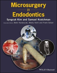
Microsurgery in Endodontics (eBook, PDF)
Redaktion: Kim, Syngcuk; Setzer, Frank; Kohli, Meetu; Karabucak, Bekir; Kratchman, Samuel
Alle Infos zum eBook verschenken
Microsurgery in Endodontics (eBook, PDF)
Redaktion: Kim, Syngcuk; Setzer, Frank; Kohli, Meetu; Karabucak, Bekir; Kratchman, Samuel
- Format: PDF
- Merkliste
- Auf die Merkliste
- Bewerten Bewerten
- Teilen
- Produkt teilen
- Produkterinnerung
- Produkterinnerung
Hier können Sie sich einloggen
Bitte loggen Sie sich zunächst in Ihr Kundenkonto ein oder registrieren Sie sich bei bücher.de, um das eBook-Abo tolino select nutzen zu können.
Microsurgery in Endodontics provides the definitive reference to endodontic microsurgery, with instructive photographs and illustrations. * Provides a definitive reference work on endodontic microsurgery * Includes contributions from pioneers and innovators in the field of microsurgical endodontics * Describes techniques for a wide range of microsurgical procedures * Includes more than 600 instructive illustrations and photographs
- Geräte: PC
- ohne Kopierschutz
- eBook Hilfe
- Größe: 167.28MB
- Microsurgery in Endodontics (eBook, ePUB)156,99 €
- Seth DelpachitraPrinciples of Dentoalveolar Extractions (eBook, PDF)114,99 €
- Impacted Third Molars (eBook, PDF)115,99 €
- U. J. MoorePrinciples of Oral and Maxillofacial Surgery (eBook, PDF)55,99 €
- Serge DibartPractical Osseous Surgery in Periodontics and Implant Dentistry (eBook, PDF)80,99 €
- Cosmesis of the Mouth, Face and Jaws (eBook, PDF)201,99 €
- Orthognathic Surgery (eBook, PDF)177,99 €
-
-
-
Dieser Download kann aus rechtlichen Gründen nur mit Rechnungsadresse in A, B, BG, CY, CZ, D, DK, EW, E, FIN, F, GR, HR, H, IRL, I, LT, L, LR, M, NL, PL, P, R, S, SLO, SK ausgeliefert werden.
- Produktdetails
- Verlag: John Wiley & Sons
- Seitenzahl: 256
- Erscheinungstermin: 26. Juli 2017
- Englisch
- ISBN-13: 9781119403654
- Artikelnr.: 48958582
- Verlag: John Wiley & Sons
- Seitenzahl: 256
- Erscheinungstermin: 26. Juli 2017
- Englisch
- ISBN-13: 9781119403654
- Artikelnr.: 48958582
- Herstellerkennzeichnung Die Herstellerinformationen sind derzeit nicht verfügbar.
Preface xv
Acknowledgements xvii
1 The Dental OperatingMicroscope 1
Frank Setzer
1.1 Benefits of the Operating Microscope 1
1.2 Key Features of Operating Microscopes 1
1.3 Customizing a Microscope 3
1.3.1 Light Source 3
1.3.2 Documentation 5
1.3.3 Individual Microscope Adjustment (Parfocaling) 5
Suggested Readings 7
2 Microsurgical Instruments 9
Seung Ho Baek and Syngcuk Kim
2.1 Examination Instruments 9
2.2 Incision and Elevation Instrument 9
2.3 Tissue Retraction Instruments 11
2.4 Osteotomy Instruments 14
2.5 Curettage Instruments 14
2.6 Inspection Instruments 15
2.7 Ultrasonic Units and Tips for Root End Preparation 17
2.8 Microplugger Instruments 20
2.9 Suturing Instruments 20
2.10 Miscellaneous Instruments 22
3 Medication-Related Osteonecrosis of the Jaw and EndodonticMicrosurgery 25
Chafic Safi and Bekir Karabucak
Suggested Readings 29
4 Indications and Contraindications 31
Bekir Karabucak and Garrett Guess
4.1 Introduction 31
4.2 Surgical Success Dependent on Ability to Perform Ideal Protocols 31
4.3 Etiology Assessment through Examination and Treatment 31
4.4 Periodontal Considerations and Surgery 33
4.5 Influential Patient Factors 33
4.6 Condition of Previous Endodontic Treatment 37
Suggested Readings 38
5 Anesthesia and Hemostasis 39
Siva Rethnam-Haug, Aleksander Iofin, and Syngcuk Kim
5.1 Armamentarium 39
5.1.1 Epinephrine 39
5.2 Presurgical Phase 40
5.2.1 Administration of Local Anesthetic 40
5.2.2 Injection Techniques 40
5.2.3 Topical Anesthesia 41
5.2.4 Additional Techniques 41
5.2.5 Maxillary Anesthesia 41
5.2.6 Mandibular Anesthesia 43
5.2.7 Bilateral Mandibular Surgery 43
5.3 Surgical Phase 44
5.3.1 Topical Hemostatic Agents 44
5.3.1.1 Epinephrine Pellets 45
5.3.1.2 Ferric Sulfate 45
5.4 Summary of Hemostatic Techniques in Endodontic Microsurgery 46
5.5 Postsurgical Phase 46
Suggested Readings 48
6 Flap Design in EndodonticMicrosurgery 49
FrancescoMaggiore and Frank Setzer
6.1 Armamentarium 49
6.1.1 Flap Outline 49
6.1.2 Papilla Management 52
6.1.3 Incision 54
6.1.4 Flap Elevation 54
6.1.5 Flap Retraction 55
Suggested Readings 56
7 Osteotomy 57
FrancescoMaggiore and Syngcuk Kim
7.1 Armamentarium 57
7.2 Osteotomy 57
7.2.1 Distinction between Bone and Root Tip 57
7.2.2 Clinical Situations for Endodontic Microsurgery 58
7.3 Intact Cortical Plate without a Radiographic Periapical Lesion 59
7.4 Intact Cortical Plate with a Periapical Lesion 60
7.5 Fenestration through the Cortical Plate Leading to the Apex 60
7.5.1 Optimal Osteotomy Size 61
7.5.2 Key Hole Osteotomy Modification 61
7.5.3 BoneWindow Technique 61
8 Root End Resection 67
Spyros Floratos, Fouad Al-Malki, and Syngcuk Kim
8.1 Armamentarium 67
8.2 Root End Resection 67
8.3 Root End Resection: Steep Bevel versus Shallow Bevel 69
Suggested Readings 72
9 Inspection of the Resected Root Surface: Importance of Isthmus 73
Spyros Floratos, Jorge Vera, Fouad Al-Malki, and Syngcuk Kim
9.1 Armamentarium 73
9.1.1 Methylene Blue Staining (MBS) 73
&nbs