40,95 €
40,95 €
inkl. MwSt.
Sofort per Download lieferbar
payback
20 °P sammeln
40,95 €
40,95 €
inkl. MwSt.
Sofort per Download lieferbar

Alle Infos zum eBook verschenken
payback
20 °P sammeln
Als Download kaufen
40,95 €
inkl. MwSt.
Sofort per Download lieferbar
payback
20 °P sammeln
Jetzt verschenken
40,95 €
inkl. MwSt.
Sofort per Download lieferbar

Alle Infos zum eBook verschenken
payback
20 °P sammeln
  • Format: PDF

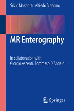
In recent years, there has been huge interest in developing new methods that offer improved accuracy for the detection of small bowel pathology, and in particular for the assessment of inflammatory bowel diseases (IBD). Cross-sectional imaging, such as CT and MR, has advantages over traditional barium fluoroscopic techniques in terms of direct visualization of the bowel wall and improved visualization of extraluminal findings and complications. This means a complete change in the diagnostic approach to the patient with IBD: from analysis of the bowel surface to direct evaluation of parietal…mehr

Produktbeschreibung
In recent years, there has been huge interest in developing new methods that offer improved accuracy for the detection of small bowel pathology, and in particular for the assessment of inflammatory bowel diseases (IBD). Cross-sectional imaging, such as CT and MR, has advantages over traditional barium fluoroscopic techniques in terms of direct visualization of the bowel wall and improved visualization of extraluminal findings and complications. This means a complete change in the diagnostic approach to the patient with IBD: from analysis of the bowel surface to direct evaluation of parietal alterations and assessment of peri- and extraintestinal involvement. The ideal imaging test is reproducible, well tolerated by patients and, above all, free of ionizing radiation. MR enterography, currently performed only in a few reference centers, meets these criteria and offers accurate diagnosis, particularly in respect of the wide spectrum of intra- and extraintestinal complications of IBD.

This book provides a thorough overview of the indications, techniques, diagnostic advantages, and limitations of MR enterography. Particular attention is paid to patient preparation in relation to the particular study type and to the potential advantages of the most up-to-date MR studies in specific cases, e.g., allergy or renal failure. A separate chapter is devoted to MR of perianal region for the detection and staging of perianal fistula, a common complication in patients with Crohn's disease. Numerous high-quality illustrations are included and help to ensure that the book will be a valuable source of information for every radiologist involved in abdominal MR imaging.


Dieser Download kann aus rechtlichen Gründen nur mit Rechnungsadresse in A, B, BG, CY, CZ, D, DK, EW, E, FIN, F, GR, HR, H, IRL, I, LT, L, LR, M, NL, PL, P, R, S, SLO, SK ausgeliefert werden.

Autorenporträt
Professor Silvio Mazziotti is Assistant Professor in Radiology at the University of Messina, Italy. He is the author or co-author of more than 130 publications in the field of diagnostic imaging, including monographs, book chapters and over 70 journal articles. Professor Mazziotti is co-author of two previous books published by Springer: I. Pandolfo and S. Mazziotti (2013) Orthopantomography and E. Neri and S. Mazziotti (2012) Il Dentascan. He acts as a reviewer for a number of leading journals.

Professor Alfredo Blandino is Chief of MRI in the Department of Radiation Oncology at the Polyclinic of the University of Messina. He is author or co-author of more than 200 publications in the field of diagnostic imaging, including books, monographs and book chapters. He has already published with Springer and has served on the editorial boards of various journals, including La radiologia medica and European Radiology.