48,95 €
48,95 €
inkl. MwSt.
Sofort per Download lieferbar
48,95 €
48,95 €
inkl. MwSt.
Sofort per Download lieferbar

Alle Infos zum eBook verschenken
Als Download kaufen
48,95 €
inkl. MwSt.
Sofort per Download lieferbar
Jetzt verschenken
48,95 €
inkl. MwSt.
Sofort per Download lieferbar

Alle Infos zum eBook verschenken
  • Format: PDF

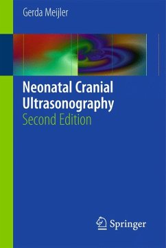
Cranial ultrasonography is the most accessible, patient friendly, and cheapest neuroimaging technique on the neonatal ward. It provides important information on brain maturation in the (preterm) neonate and enables the detection of frequently occurring brain anomalies in both preterm and full term neonates. Furthermore, it can be repeated as often as is necessary.
In this second edition of Neonatal Cranial Ultrasonography, the focus is on the basics of the technique, from patient preparation through to screening strategies and the classification of abnormalities. Many new ultrasound images
…mehr

  • Geräte: PC
  • ohne Kopierschutz
  • eBook Hilfe
  • Größe: 11.33MB
Produktbeschreibung
Cranial ultrasonography is the most accessible, patient friendly, and cheapest neuroimaging technique on the neonatal ward. It provides important information on brain maturation in the (preterm) neonate and enables the detection of frequently occurring brain anomalies in both preterm and full term neonates. Furthermore, it can be repeated as often as is necessary.

In this second edition of Neonatal Cranial Ultrasonography, the focus is on the basics of the technique, from patient preparation through to screening strategies and the classification of abnormalities. Many new ultrasound images have been included to reflect the improvements in image quality since the first edition. Essential information is provided about both the procedure itself and the normal ultrasound anatomy. Standard technique using the anterior fontanelle as the acoustic window is described and illustrated, but emphasis is also placed on the value of supplementary acoustic windows. The compact design of the book makes it an ideal and handy reference that will guide the novice but also provide useful information for the more experienced practitioner.


Dieser Download kann aus rechtlichen Gründen nur mit Rechnungsadresse in A, B, BG, CY, CZ, D, DK, EW, E, FIN, F, GR, HR, H, IRL, I, LT, L, LR, M, NL, PL, P, R, S, SLO, SK ausgeliefert werden.

Autorenporträt
Gerda Meijler is pediatrician-neonatologist. She studied medicine at the University of Amsterdam and the Academic Medical Center. She did her pediatric and neonatology training respectively at the Wilhelmina Children's Hospital, Utrecht and the Academic Medical Center, Amsterdam. After being a staff-neonatologist at the VU Medical Center, Amsterdam, where she did her PhD study on brain imaging in preterm neonates, she moved to the Leiden University Medical Center in 1999. Since then her special interest is neonatal neurology and neuro-imaging. She is the principal investigator of the Leiden neonatal neuro-imaging group. This group is actively involved in research in this field and is known for its high quality neuro-imaging (both cranial ultrasound and MRI). Together with this group Gerda has further improved neuro-imaging techniques, both for clinical and research purposes and has introduced the routine use of alternative acoustic windows in cranial ultrasonography. This has resulted in optimizing ultrasound imaging of the neonatal cerebellum. Gerda teaches neonatal neuro-imaging, both in the Netherlands and abroad at several courses and congresses. Her research now mainly focuses on imaging of the neonatal cerebellum, brain imaging and injury in the preterm neonate and on applying advanced imaging techniques in neonates.

After the summer of 2011 she will move to Toronto, where she will continue her work on neonatal neurology and neuro-imaging at the neonatal units of the Hospital for Sick Children and the Mount Sinaï Hospital.