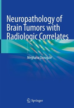
This highly illustrated book explores the pathological and radiological diagnosis of various brain tumors. Featuring 900 high-quality colored images, it covers MR images, intra-operative squash cytology, histopathology and immunohistochemistry microphotographs of various brain and spine tumors, including differential diagnosis, as well as the molecular diagnosis and prognosis of each tumor. The book also presents case studies of typical and rare presentations, and introduces readers to a new procedure for intra-operative cytology: the modified fields stain, which stains the slide within 2 minutes, allowing quick, accurate reporting.
This book uses concise text and a consistent point-wise format that makes reading and reviewing easy. The radiological and pathological correlates of brain and spine tumors serve as a ready-reference resource for residents, surgical and neuropathologists, neuroradiologists, neurosurgeons, neuro-oncologists and research scientists.
Dieser Download kann aus rechtlichen Gründen nur mit Rechnungsadresse in A, B, BG, CY, CZ, D, DK, EW, E, FIN, F, GR, HR, H, IRL, I, LT, L, LR, M, NL, PL, P, R, S, SLO, SK ausgeliefert werden.

