Ophthalmological Imaging and Applications (eBook, PDF)
Redaktion: Ng, E. Y. K.; Suri, Jasjit S.; Rangayyan, Rangaraj M.; Acharya, U. Rajendra
Alle Infos zum eBook verschenken
Ophthalmological Imaging and Applications (eBook, PDF)
Redaktion: Ng, E. Y. K.; Suri, Jasjit S.; Rangayyan, Rangaraj M.; Acharya, U. Rajendra
- Format: PDF
- Merkliste
- Auf die Merkliste
- Bewerten Bewerten
- Teilen
- Produkt teilen
- Produkterinnerung
- Produkterinnerung
Hier können Sie sich einloggen
Bitte loggen Sie sich zunächst in Ihr Kundenkonto ein oder registrieren Sie sich bei bücher.de, um das eBook-Abo tolino select nutzen zu können.
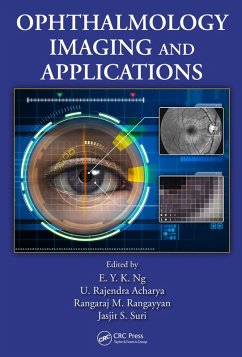
This book covers several aspects of multimodal ophthalmological imaging and applications. Edited by and featuring contributions from world-class researchers, the text offers a unified work of the latest eye imaging and modeling techniques proposed and applied to ophthalmologic problems. It focuses on theory, basic principles, and results derived from research. Information is presented in an accessible manner to appeal to a wide audience of students, researchers, and practitioners. This volume is intended to be a companion to Image Analysis and Modeling in Ophthalmology.
- Geräte: PC
- mit Kopierschutz
- eBook Hilfe
- Größe: 61.63MB
- Samuel C. MorrisCancer Risk Assessment (eBook, PDF)65,95 €
- Arkadiusz SitekStatistical Computing in Nuclear Imaging (eBook, PDF)48,95 €
- Image Analysis and Modeling in Ophthalmology (eBook, PDF)96,95 €
- Nuclear Medicine Physics (eBook, PDF)61,95 €
- John L. SemmlowBiosignal and Medical Image Processing (eBook, PDF)141,95 €
- Luca PanconiMethods for Imaging Cell Membranes (eBook, PDF)52,95 €
- Wolfgang BirkfellnerApplied Medical Image Processing (eBook, PDF)75,95 €
-
-
-
Dieser Download kann aus rechtlichen Gründen nur mit Rechnungsadresse in A, B, BG, CY, CZ, D, DK, EW, E, FIN, F, GR, HR, H, IRL, I, LT, L, LR, M, NL, PL, P, R, S, SLO, SK ausgeliefert werden.
- Produktdetails
- Verlag: Taylor & Francis
- Seitenzahl: 529
- Erscheinungstermin: 5. Mai 2014
- Englisch
- ISBN-13: 9781466559158
- Artikelnr.: 41068574
- Verlag: Taylor & Francis
- Seitenzahl: 529
- Erscheinungstermin: 5. Mai 2014
- Englisch
- ISBN-13: 9781466559158
- Artikelnr.: 41068574
- Herstellerkennzeichnung Die Herstellerinformationen sind derzeit nicht verfügbar.
the Major Temporal Arcade in Retinal Fundus Images. Application of Higher
Order Spectra Cumulants for Diabetic Retinopathy Detection using Digital
Fundus Images. Quality Measures for Retinal Images. Graph Search Retinal
Vessel Tracking. Fundus Autofluorescence Imaging. The Needs/Requirements
and Design of Imaging and Image Processing Methods for Ophthalmology in the
Indian Context. The Application of Ocular Fundus Photography and
Angiography. Optic Nerve Analysis and Imaging in Relation to Glaucoma.
Imaging of the Eye after Glaucoma Surgery. Confocal Microscopy of Cornea.
Corneal Topography and Tomography. Automatic Analysis of Scanning Laser
Ophthalmoscope Sequences for Arteriovenous Passage Time Measurement.
Optical Coherence Tomography. The Role of Optical Coherence Tomography on
Imaging of the Ocular Surface. Anterior Segment Imaging of Anterior Segment
Optical Coherence Tomography. Cyst Detection in OCT Images for Pathology
Characterization. Scanning Laser Ophthalmoscope Fundus Perimetry. In Vivo
Confocal Microscopy. Biomechanical Modeling of Blood Vessels for
Interpretation of Tortuosity Estimates. Hybrid Finite Element Simulation
for Bioheat Transfer in Human Eye. Effects of Electromagnetic Fields on
Specific Absorption Rate and Heat Transfer in the Human Eye. Dry-Eye
Characterization by Analysis of Tear Film Images. Thermography and the Eye.