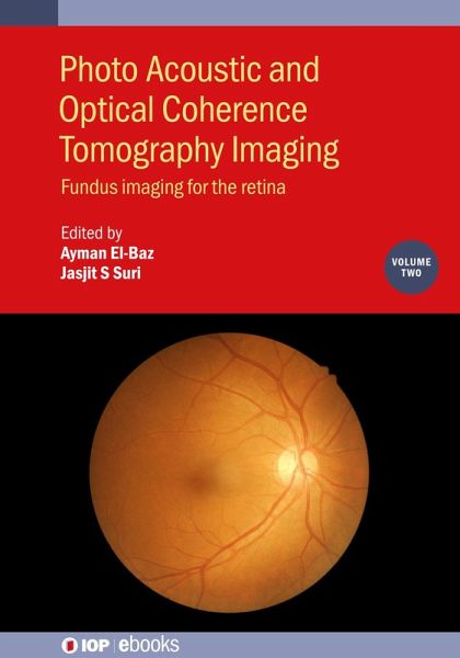
Photo Acoustic and Optical Coherence Tomography Imaging, Volume 2 (eBook, ePUB)
Fundus imaging for the retina
Redaktion: El-Baz, Ayman; Suri, Jasjit S
Versandkostenfrei!
Sofort per Download lieferbar
79,95 €
inkl. MwSt.
Weitere Ausgaben:

PAYBACK Punkte
40 °P sammeln!
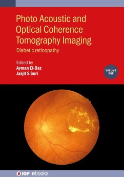
This book covers the state-of-the-art techniques of fundus imaging for the diagnosis of retinal diseases. It is part of a three-volume work that describes the latest imaging techniques in which to bring optical coherence tomography (OCT), fundus Imaging and optical coherence tomography angiography (OCTA) to accurately facilitate the diagnosis of retinal diseases. Clinical disorders of the retina have been attracting the attention of researchers, aiming at reducing the blindness rate. This includes uveitis, diabetic retinopathy, macular edema, endophthalmitis, proliferative retinopathy, age-rel...
This book covers the state-of-the-art techniques of fundus imaging for the diagnosis of retinal diseases. It is part of a three-volume work that describes the latest imaging techniques in which to bring optical coherence tomography (OCT), fundus Imaging and optical coherence tomography angiography (OCTA) to accurately facilitate the diagnosis of retinal diseases. Clinical disorders of the retina have been attracting the attention of researchers, aiming at reducing the blindness rate. This includes uveitis, diabetic retinopathy, macular edema, endophthalmitis, proliferative retinopathy, age-related macular degeneration and glaucoma. Treatment is significantly dependent on having early and accurate diagnosis, which can be significantly improved by employing the techniques described in the book.
Key Features
Key Features
- Provides a comprehensive overview of all pertinent topics related to fundus imaging techniques, applicable to diagnosis of eye disorders
- Offers a unique coverage of Neural Networks in distinguishing eye diseases
- Machine learning techniques are presented in detail throughout
- Many of the chapter contributors are world-class researchers
- Extensive references will be provided at the end of each chapter to enhance further study
Dieser Download kann aus rechtlichen Gründen nur mit Rechnungsadresse in A, D ausgeliefert werden.


