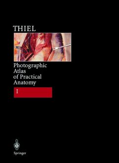
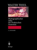
Professor Walter Thiels brilliant photographs are unique. They revolutionise macroscopic surgery because - due to a new preservation technique developed by the author himself - all tissues retain their living colour, consistency and position. This new technique, Thiels exceptional abilities as a photographer and the filigree dissections add up to vivid, almost artistic illustrations of astonishing depth and clarity. Apart from the topographical anatomy of the abdomen and lower extremities, Part I illustrates the most important punctures of joints and many surgical approaches. Thus this atlas is not only of interest to anatomists and pathologists but particularly to surgeons and orthopaedic surgeons - in fact all doctors requiring a 3D presentation of human anatomy.
Dieser Download kann aus rechtlichen Gründen nur mit Rechnungsadresse in A, B, BG, CY, CZ, D, DK, EW, E, FIN, F, GR, HR, H, IRL, I, LT, L, LR, M, NL, PL, P, R, S, SLO, SK ausgeliefert werden.
Hinweis: Dieser Artikel kann nur an eine deutsche Lieferadresse ausgeliefert werden.

