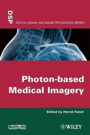
Photon-based Medical Imagery (eBook, ePUB)
Redaktion: Fanet, Hervé
144,99 €
144,99 €
inkl. MwSt.
Sofort per Download lieferbar
0 °P sammeln
144,99 €
Als Download kaufen
144,99 €
inkl. MwSt.
Sofort per Download lieferbar
0 °P sammeln
Jetzt verschenken
Alle Infos zum eBook verschenken
144,99 €
inkl. MwSt.
Sofort per Download lieferbar
Alle Infos zum eBook verschenken
0 °P sammeln
Photon-based Medical Imagery (eBook, ePUB)
Redaktion: Fanet, Hervé
- Format: ePub
- Merkliste
- Auf die Merkliste
- Bewerten Bewerten
- Teilen
- Produkt teilen
- Produkterinnerung
- Produkterinnerung
Bitte loggen Sie sich zunächst in Ihr Kundenkonto ein oder registrieren Sie sich bei
bücher.de, um das eBook-Abo tolino select nutzen zu können.
Hier können Sie sich einloggen
Sie sind bereits eingeloggt. Klicken Sie auf 2. tolino select Abo, um fortzufahren.
Bitte loggen Sie sich zunächst in Ihr Kundenkonto ein oder registrieren Sie sich bei bücher.de, um das eBook-Abo tolino select nutzen zu können.
This book describes the different principles and equipments used in medical imaging. Importance of medical imaging for diagnostic is strongly increasing and it is now necessary to have a good knowledge of the different physical possible principles. Researchers, clinicians, engineers and professionals in this area, along with postgraduate students in the signal and image processing field, will find this book of great interest.
- Geräte: eReader
- mit Kopierschutz
- eBook Hilfe
- Größe: 3.29MB
Andere Kunden interessierten sich auch für
- Mark HaidekkerAdvanced Biomedical Image Analysis (eBook, ePUB)148,99 €
- Photon-based Medical Imagery (eBook, PDF)144,99 €
- H. K. HuangPACS-Based Multimedia Imaging Informatics (eBook, ePUB)169,99 €
- Advances in Optical Imaging for Clinical Medicine (eBook, ePUB)160,99 €
- Adam de la ZerdaBiochips and Medical Imaging (eBook, ePUB)138,99 €
- Atam P. DhawanMedical Image Analysis (eBook, ePUB)129,99 €
- Biomedical Imaging (eBook, ePUB)120,99 €
-
-
-
This book describes the different principles and equipments used in medical imaging. Importance of medical imaging for diagnostic is strongly increasing and it is now necessary to have a good knowledge of the different physical possible principles. Researchers, clinicians, engineers and professionals in this area, along with postgraduate students in the signal and image processing field, will find this book of great interest.
Dieser Download kann aus rechtlichen Gründen nur mit Rechnungsadresse in A, B, BG, CY, CZ, D, DK, EW, E, FIN, F, GR, HR, H, IRL, I, LT, L, LR, M, NL, PL, P, R, S, SLO, SK ausgeliefert werden.
Produktdetails
- Produktdetails
- Verlag: Wiley
- Seitenzahl: 330
- Erscheinungstermin: 4. Februar 2013
- Englisch
- ISBN-13: 9781118601228
- Artikelnr.: 37486212
- Verlag: Wiley
- Seitenzahl: 330
- Erscheinungstermin: 4. Februar 2013
- Englisch
- ISBN-13: 9781118601228
- Artikelnr.: 37486212
- Herstellerkennzeichnung Die Herstellerinformationen sind derzeit nicht verfügbar.
Hervé Fanet, Research engineer at CEA Leti. He is in charge of scientific cooperation and training activities within the MINATEC (micro and nanotechnology) in Grenoble.
Foreword xi
Guy FRIJA
Chapter 1. Interactions between Radiation and Matter: Consequences for
Detection and Medical Imaging 1
Jean-Pierre MOY
1.1. The limits of imaging using light 1
1.2. Imaging with other types of radiation 2
1.3. X-rays: their interaction with matter 3
1.4. Radiological imaging relies on the X-ray-matter interaction 19
1.5. Consequences of interaction modes on detection 22
1.6. Conclusion 33
1.7. Bibliography 33
Chapter 2. Detectors for Medical Imaging 35
Hervé FANET
2.1. Radiation-matter interaction and signal formation 36
2.2. Flux, energy, time and position measurements 64
2.3. Semi-conductor detectors 86
2.4. Scintillation and measurement channel 102
2.5. Pixel detectors 116
2.6. Bibliography 121
Chapter 3. Quantitative Digital Radiography Image Processing 123
Jean RINKEL and Jean-Marc DINTEN
3.1. Introduction to flat-panel sensors 123
3.2. Relation between physical quantities and radiographic acquisition 129
3.3. Access to linear attenuation coefficients from the attenuation image
133
3.4. Access to physical dimensions by combining several X-rays of a flat
sensor 149
3.5. Conclusion 158
3.6. Bibliography 159
Chapter 4. X-Ray Tomography 161
Françoise PEYRIN and Philippe DOUEK
4.1. Introduction 161
4.2. Principle of the first acquisition systems 162
4.3. Physical aspects and the direct problem 165
4.4. Principle of tomographic image reconstruction 169
4.5. Evolution of X-ray scanners and reconstruction algorithms 181
4.6. Examples of clinical applications 189
4.7. From tomography to micro-tomography 198
4.8. Conclusion 200
4.9. Bibliography 201
Chapter 5. Positron-Emission Tomography: Principles and Applications 207
Régine TRÉBOSSEN
5.1. Introduction 208
5.2. PET: principle and performance 210
5.3. PET systems 218
5.4. PET for cancer staging 223
5.5. Conclusion 225
5.6. Bibliography 226
Chapter 6. Single Photon Imaging 229
Irène BUVAT
6.1. Introduction 229
6.2. Overview of single photon imaging 230
6.3. Conventional detection systems in single photon imaging: the
scintillation gamma camera 233
6.4. Innovative systems: semiconductor detectors 240
6.5. Tomographic reconstruction and corrections 241
6.6. Hybrid detectors 258
6.7. Applications 259
6.8. Future developments 261
6.9. Conclusion 262
6.10. Bibliography 262
Chapter 7. Optical Imaging 267
Anabela DA SILVA
7.1. Introduction 267
7.2. Physics of luminous propagation in biological tissue 268
7.3. Different optical imaging techniques for different applications 289
7.4. Conclusion 312
7.5. Bibliography 313
List of Authors 325
Index 327