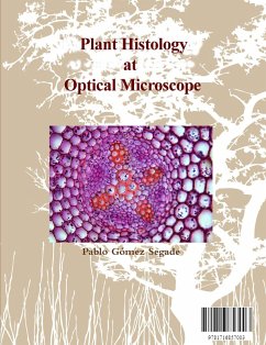
The main body of this book is a set of 91 color photomicrographs, most of them at 600 dpi and obtained with an Olympus microscope from the Department of Animal Biology (before called Cytology and Histology) of the Faculty of Biology of the University of Santiago of Compostela. It includes photomicrographs from the cyanobacteria to the higher plants, passing through fungi, mosses, liverworts and ferns. In the plants we study all kinds of tissues as well as sections of different parts such as the root, stem, leaf and the female and male parts of the flower. The book includes at the end an evaluation section consisting of clippings of the main photos, with related questions. Also included is an annex of 48 micrographs made with the Motic DM-B1 microscope on bacteria, single-celled algae, fungi and complementary sections of plant structures. The work is directed both to students and teachers of "Plant Histology" or "Botany" of university degrees such as those offered in faculties such as Biology, Pharmacy, Primary School, Agricultural Engineering ... This work is of interest to the teachers of Secondary School who teach "Biology and Geology" of fourth year of the Compulsary Secondary Education. And "Biology and Geology" of the first year of the baccalaureate of Sciences of the Nature and the Health. In general, the book can be useful to teachers of other levels of education related to the world of plants and vegetables. The work is for me an old aim since I had the pleasure of teaching laboratory practices of "Histology" during my doctoral thesis in the Faculty of Biology of the University of Santiago of Compostela. My main wish is that the spectacular colours and designs of the microscopic interior world of plants and other groups some way related should be shared and enjoyed by all.
Dieser Download kann aus rechtlichen Gründen nur mit Rechnungsadresse in A, B, BG, CY, CZ, D, DK, EW, E, FIN, F, GR, HR, H, IRL, I, LT, L, LR, M, NL, PL, P, R, S, SLO, SK ausgeliefert werden.

