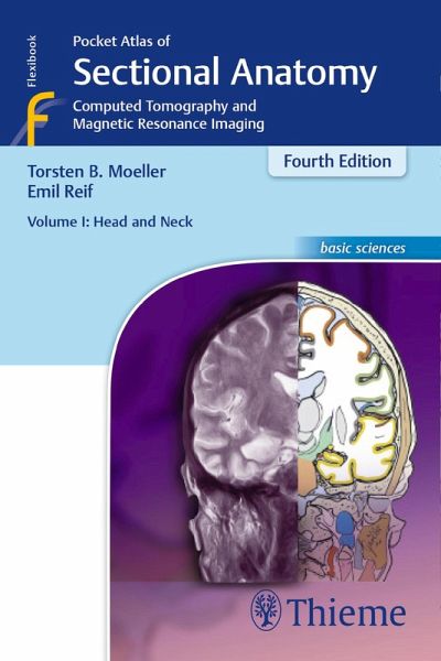
Pocket Atlas of Sectional Anatomy, Volume I: Head and Neck (eBook, ePUB)
Computed Tomography and Magnetic Resonance Imaging
Versandkostenfrei!
Sofort per Download lieferbar
Statt: 51,00 €**
36,95 €
inkl. MwSt.
**Preis der gedruckten Ausgabe (Broschiertes Buch)
Alle Infos zum eBook verschenkenWeitere Ausgaben:

PAYBACK Punkte
18 °P sammeln!
This comprehensive, easy-to-consult pocket atlas is renowned for its superb illustrations and ability to depict sectional anatomy in every plane. Together with its two companion volumes, it provides a highly specialized navigational tool for all clinicians who need to master radiologic anatomy and accurately interpret CT and MR images.Special features of Pocket Atlas of Sectional Anatomy:Didactic organization in two-page units, with high-quality radiographs on one side and brilliant, full-color diagrams on the other Hundreds of high-resolution CT and MR images made with the latest generation o...
This comprehensive, easy-to-consult pocket atlas is renowned for its superb illustrations and ability to depict sectional anatomy in every plane. Together with its two companion volumes, it provides a highly specialized navigational tool for all clinicians who need to master radiologic anatomy and accurately interpret CT and MR images.
Special features of Pocket Atlas of Sectional Anatomy:
Updates for the 4th edition of Volume I:
Compact, easy-to-use, highly visual, and designed for quick recall, this book is ideal for use in both the clinical and study settings.
Special features of Pocket Atlas of Sectional Anatomy:
- Didactic organization in two-page units, with high-quality radiographs on one side and brilliant, full-color diagrams on the other
- Hundreds of high-resolution CT and MR images made with the latest generation of scanners (e.g., 3T MRI, 64-slice CT)
- Consistent color coding, making it easy to identify similar structures across several slices
- Concise, easy-to-read labeling of all figures
Updates for the 4th edition of Volume I:
- New cranial CT imaging sequences of the axial and coronal temporal bone
- Expanded MR section, with all new 3T MR images of the temporal lobe and hippocampus, basilar artery, cranial nerves, cavernous sinus, and more
- New arterial MR angiography sequences of the neck and additional larynx images
Compact, easy-to-use, highly visual, and designed for quick recall, this book is ideal for use in both the clinical and study settings.
Dieser Download kann aus rechtlichen Gründen nur mit Rechnungsadresse in A, B, BG, CY, CZ, D, DK, EW, E, FIN, F, GR, HR, H, IRL, I, LT, L, LR, M, NL, PL, P, R, S, SLO, SK ausgeliefert werden.


