Sie sind bereits eingeloggt. Klicken Sie auf 2. tolino select Abo, um fortzufahren.
Bitte loggen Sie sich zunächst in Ihr Kundenkonto ein oder registrieren Sie sich bei bücher.de, um das eBook-Abo tolino select nutzen zu können.
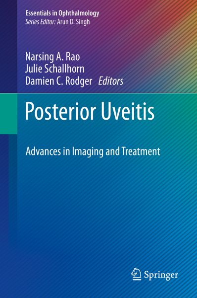
This comprehensive text provides readers with an in-depth examination of posterior uveitis, and expert instruction on diagnosis, imaging techniques and treatments that are being reshaped by advancements in the field. Posterior Uveitis: Advances in Imaging and Treatment focuses on the ocular imaging modalities used in the diagnosis of various uveitis and intraocular inflammation entities resulting from infectious and non-infectious etiologies. Each topic is succinctly presented by experts in the field of intraocular inflammation and ocular imaging and starts with salient clinical features,…mehr
This comprehensive text provides readers with an in-depth examination of posterior uveitis, and expert instruction on diagnosis, imaging techniques and treatments that are being reshaped by advancements in the field. Posterior Uveitis: Advances in Imaging and Treatment focuses on the ocular imaging modalities used in the diagnosis of various uveitis and intraocular inflammation entities resulting from infectious and non-infectious etiologies. Each topic is succinctly presented by experts in the field of intraocular inflammation and ocular imaging and starts with salient clinical features, differential diagnosis and specific treatment, and concludes with in-depth and relevant clinical imaging findings. The book opens by touring a multitude of infectious and non-infectious uveitidies and explores how advances are aiding our diagnosis and treatment. The second half will delve into established and emerging therapeutics, including advances in drug delivery. Evolvingtreatments for recalcitrant uveitis are discussed, including the newer biological agents, and each chapter includes ample illustrations and several tables for readers to comprehend with ease the inflammatory disorders and to interpret the imaging changes in various uveitis entities.
Dieser Download kann aus rechtlichen Gründen nur mit Rechnungsadresse in A, B, BG, CY, CZ, D, DK, EW, E, FIN, F, GR, HR, H, IRL, I, LT, L, LR, M, NL, PL, P, R, S, SLO, SK ausgeliefert werden.
Die Herstellerinformationen sind derzeit nicht verfügbar.
Autorenporträt
Narsing A. Rao, MD, is professor of ophthalmology and director of the Uveitis Service and the Ophthalmic Pathology Laboratory at USC. He is involved in the treatment of inflammatory ocular diseases affecting the uveal tract, vitreous, retina, and sclera. Dr. Rao has published over 440 peer-reviewed papers in clinical and basic science findings and has received numerous honors and awards for his research endeavors on ocular inflammatory diseases from Research to Prevent Blindness and the National Institute of Health. He delivered several named lectures, both national and international, including visiting professorship at Harvard University, Johns Hopkins University, Yale University, University of Paris, France, Moorefields Eye Institute, London, University of Rome, Italy, Tokyo Medical University, Kyushu University in Fukoka, Japan, and University of Sydney. For his outstanding contributions to ophthalmology, he was awarded the Lorenz E. Zimmerman medal from American Academy of Ophthalmology, the Ramon L. Font medal from Pan American Society of Ophthalmology, the International Ocular Inflammation Society Award, and received an award from European Vision and Eye Research Association in 2006. Dr. Rao was also the recipient of the prestigious Bietti medal from International Council of Ophthalmology. Narsing A. Rao, MD Department of Ophthalmology USC-Roski Eye Institute University of Southern California Los Angeles, CA USA Damien C. Rodger, MD, PhD, worked as a co-op intern at the NASA Jet Propulsion Laboratory on attitude control microsystems for unmanned space exploration while completing his Bachelor's in electrical engineering from Cornell University in 2000. Dr. Rodger then earned his Medical Degree from the Keck School of Medicine of USC and his PhD in bioengineering from the California Institute of Technology. Heis Assistant Professor of Clinical Ophthalmology and Research Assistant Professor of Biomedical Engineering at USC. He was awarded the Heed Fellowship for the 2013-2014 year and the Ronald G. Michels Fellowship in Vitreoretinal Surgery for the 2014-2015 year. He specializes in medical and surgical retina, as well as in uveitis and ocular inflammation. Damien C. Rodger MD, PhD Southern California Permanente Medical Group Department of Ophthalmology Kaiser Permanente Los Angeles Medical Center Los Angeles, CA USA Julie M. Schallhorn, MD, MS, is Assistant Professor of Clinical Ophthalmology at Keck Medicine of USC. She specializes in the surgical treatment of cataracts, corneal transplantation, lens implantation, and anterior segment disease. Her research interests include optical coherence tomography and its applications to the anterior segment, new methods of corneal transplantation, refractive surgery and ocular inflammatory diseases. Julie Schallhorn, MD MS Department of Ophthalmology F.I. Proctor Foundation University of California, San Francisco San Francisco, CA USA
Inhaltsangabe
A. Introduction.- 1. Current imaging modalities in Diagnosis and Management of Intraocular Inflammation.- Phillip Phuc Le (Anterior segment OCT, OCT imaging of Retina, OCT-EDI imaging of Retina and Choroid, Wide field fluorescein and ICG angiography, Auto-fluorescence, OCT angiography and Evolving imaging modalities).-B. Non-Infectious Posterior and Pan Uveitis.- 2. Sarcoidosis: Padmamalini Mahendradas (Introduction, main clinical features, differential diagnosis and treatment, wide field color fundus photo, fluorescein angiography and ICG angiography, OCT, Autofluorescence, Evolving imaging modalities (e.g. OCT angiography and others), Response to treatment imaging) 3. Vogt-Koyanagi-Harada disease and Sympathetic Ophthalmia: Jeffrey J. Tan and Narsing.- Rao (Introduction, main clinical, features, differential diagnosis and treatment, Wide field color fundus photo, fluorescein angiography and ICG angiography, OCT, Autofluorescence, Evolving imaging modalities (e.g. OCT angiography and others), Response to treatment imaging) .- 4. Multifocal Choroiditis/ Serpiginous Choroiditis and related entities: Hossein Nazari.- Khanamiri and Narsing Rao (Introduction, clinical features, differential diagnosis and treatment, Wide field color fundus photo, fluorescein angiography and ICG angiography, OCT, Autofluorescence, Evolving imaging modalities (e.g. OCT angiography and others), Response to treatment imaging5. Ocular Toxoplasmosis: Daniel Vitor Vasconcelos Santos (Introduction, clinical features, differential diagnosis and treatment, Wide field color fundus photo, fluorescein angiography and ICG angiography, OCT, Autofluorescence, Evolving imaging modalities (e.g. OCT angiography and others), Response to treatment imaging) .-6. Syphilis: Thomas Albini (Introduction, clinical features, differential diagnosis and treatment, Wide field color fundus photo, fluorescein angiography and ICG angiography, OCT, Autofluorescence, Evolving imaging modalities (e.g. OCT angiography and others), Response to treatment imaging) .- 7. Intraocular Tuberculosis: Soumyava Basu (Introduction, clinical features, differential diagnosis and treatment, Wide field color fundus photo, fluorescein angiography and ICG angiography, OCT, Autofluorescence, Evolving imaging modalities (e.g. OCT angiography and others), Response to treatment imaging) .-<8. Viral retinitis (HSV/VZV/CMV/PORN/Ebola): Ann-Marie Lobo (Introduction, main clinical features, differential diagnosis and treatment, Wide field color fundus photo, fluorescein angiography and ICG angiography, OCT, Autofluorescence, Evolving imaging modalities (e.g. OCT angiography and others), Response to treatment imaging) .-9. Masquerade syndromes: Damien Rodger (Wide field color fundus photo, fluorescein angiography and ICG angiography, OCT, Autofluorescence, Evolving imaging modalities (e.g. OCT angiography and others), Response to treatment imaging) .-D. Treatment of Non-Infectious Uveitis: 10. Corticosteroids: systemic and local: Ashleigh Levinson.-11. Immunomodulatory agents and Biologicals: John Gonzales and Nisha Acharya.-12. Evolving treatments: Julie Schallhorn
Rezensionen
"This book is a very worthwhile endeavor, indeed, as uveitis accounts for a significant proportion of preventable blindness despite its relative rarity as a condition in the field of ophthalmology. This book is written for ophthalmologists who wish to increase their knowledge about uveitis. As the field is constantly growing, ophthalmologists do need a nearly constant stream of updates, and books such as this fulfill that need." (Diana V Do, Doody's Book Reviews, July 12, 2019)
Es gelten unsere Allgemeinen Geschäftsbedingungen: www.buecher.de/agb
Impressum
www.buecher.de ist ein Internetauftritt der buecher.de internetstores GmbH
Geschäftsführung: Monica Sawhney | Roland Kölbl | Günter Hilger
Sitz der Gesellschaft: Batheyer Straße 115 - 117, 58099 Hagen
Postanschrift: Bürgermeister-Wegele-Str. 12, 86167 Augsburg
Amtsgericht Hagen HRB 13257
Steuernummer: 321/5800/1497
USt-IdNr: DE450055826