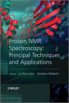
Protein NMR Spectroscopy (eBook, ePUB)
Practical Techniques and Applications
Redaktion: Lian, Lu-Yun; Roberts, Gordon
91,99 €
91,99 €
inkl. MwSt.
Sofort per Download lieferbar

0 °P sammeln
91,99 €
Als Download kaufen

91,99 €
inkl. MwSt.
Sofort per Download lieferbar

0 °P sammeln
Jetzt verschenken
Alle Infos zum eBook verschenken
91,99 €
inkl. MwSt.
Sofort per Download lieferbar
Alle Infos zum eBook verschenken

0 °P sammeln
Protein NMR Spectroscopy (eBook, ePUB)
Practical Techniques and Applications
Redaktion: Lian, Lu-Yun; Roberts, Gordon
- Format: ePub
- Merkliste
- Auf die Merkliste
- Bewerten Bewerten
- Teilen
- Produkt teilen
- Produkterinnerung
- Produkterinnerung

Bitte loggen Sie sich zunächst in Ihr Kundenkonto ein oder registrieren Sie sich bei
bücher.de, um das eBook-Abo tolino select nutzen zu können.
Hier können Sie sich einloggen
Hier können Sie sich einloggen
Sie sind bereits eingeloggt. Klicken Sie auf 2. tolino select Abo, um fortzufahren.

Bitte loggen Sie sich zunächst in Ihr Kundenkonto ein oder registrieren Sie sich bei bücher.de, um das eBook-Abo tolino select nutzen zu können.
Nuclear Magnetic Resonance (NMR) spectroscopy, a physical phenomenon based upon the magnetic properties of certain atomic nuclei, has found a wide range of applications in life sciences over recent decades. This up-to-date volume covers NMR techniques and their application to proteins, with a focus on practical details. Providing newcomers to NMR with practical guidance to carry out successful experiments with proteins and analyze the resulting spectra, those familiar with the chemical applications of NMR will also find it useful in understanding the special requirements of protein NMR.
- Geräte: eReader
- mit Kopierschutz
- eBook Hilfe
- Größe: 6.65MB
Andere Kunden interessierten sich auch für
![Applied NMR Spectroscopy for Chemists and Life Scientists (eBook, ePUB) Applied NMR Spectroscopy for Chemists and Life Scientists (eBook, ePUB)]() Oliver ZerbeApplied NMR Spectroscopy for Chemists and Life Scientists (eBook, ePUB)58,99 €
Oliver ZerbeApplied NMR Spectroscopy for Chemists and Life Scientists (eBook, ePUB)58,99 €![Spin Dynamics (eBook, ePUB) Spin Dynamics (eBook, ePUB)]() Malcolm H. LevittSpin Dynamics (eBook, ePUB)52,99 €
Malcolm H. LevittSpin Dynamics (eBook, ePUB)52,99 €![Protein NMR Spectroscopy (eBook, PDF) Protein NMR Spectroscopy (eBook, PDF)]() Protein NMR Spectroscopy (eBook, PDF)91,99 €
Protein NMR Spectroscopy (eBook, PDF)91,99 €![Understanding NMR Spectroscopy (eBook, ePUB) Understanding NMR Spectroscopy (eBook, ePUB)]() James KeelerUnderstanding NMR Spectroscopy (eBook, ePUB)39,99 €
James KeelerUnderstanding NMR Spectroscopy (eBook, ePUB)39,99 €![A Primer of NMR Theory with Calculations in Mathematica (eBook, ePUB) A Primer of NMR Theory with Calculations in Mathematica (eBook, ePUB)]() Alan J. BenesiA Primer of NMR Theory with Calculations in Mathematica (eBook, ePUB)64,99 €
Alan J. BenesiA Primer of NMR Theory with Calculations in Mathematica (eBook, ePUB)64,99 €![NMR of Quadrupolar Nuclei in Solid Materials (eBook, ePUB) NMR of Quadrupolar Nuclei in Solid Materials (eBook, ePUB)]() Roderick E. WasylishenNMR of Quadrupolar Nuclei in Solid Materials (eBook, ePUB)147,99 €
Roderick E. WasylishenNMR of Quadrupolar Nuclei in Solid Materials (eBook, ePUB)147,99 €![Medical Imaging Based on Magnetic Fields and Ultrasounds (eBook, ePUB) Medical Imaging Based on Magnetic Fields and Ultrasounds (eBook, ePUB)]() Medical Imaging Based on Magnetic Fields and Ultrasounds (eBook, ePUB)139,99 €
Medical Imaging Based on Magnetic Fields and Ultrasounds (eBook, ePUB)139,99 €-
-
-
Nuclear Magnetic Resonance (NMR) spectroscopy, a physical phenomenon based upon the magnetic properties of certain atomic nuclei, has found a wide range of applications in life sciences over recent decades. This up-to-date volume covers NMR techniques and their application to proteins, with a focus on practical details. Providing newcomers to NMR with practical guidance to carry out successful experiments with proteins and analyze the resulting spectra, those familiar with the chemical applications of NMR will also find it useful in understanding the special requirements of protein NMR.
Dieser Download kann aus rechtlichen Gründen nur mit Rechnungsadresse in D ausgeliefert werden.
Produktdetails
- Produktdetails
- Verlag: Wiley
- Erscheinungstermin: 9. Juni 2011
- Englisch
- ISBN-13: 9781119972822
- Artikelnr.: 37342564
- Verlag: Wiley
- Erscheinungstermin: 9. Juni 2011
- Englisch
- ISBN-13: 9781119972822
- Artikelnr.: 37342564
- Herstellerkennzeichnung Die Herstellerinformationen sind derzeit nicht verfügbar.
Professor Gordon Roberts is Head of the School of Biological Sciences, University of Leicester. Dr Christina Redfield is Reader, Oxford Centre for Molecular Sciences, University of Oxford.
List of Contributors xiii
Introduction 1
Lu-Yun Lian and Gordon Roberts
References 4
1 Sample Preparation, Data Collection and Processing 5
Frederick W. Muskett
1.1 Introduction 5
1.2 Sample Preparation 5
1.2.1 Initial Considerations 6
1.2.2 Additives 7
1.2.3 Sample Conditions 7
1.2.4 Special Cases 8
1.2.5 NMR Sample Tubes 9
1.2.5.1 3 mm Tubes 10
1.3 Data Collection 11
1.3.1 Locking 11
1.3.2 Tuning 11
1.3.3 Shimming 12
1.3.4 Calibrating Pulses 13
1.3.5 Acquisition Parameters 14
1.3.6 Fast Acquisition Methods 16
1.4 Data Processing 17
References 20
2 Isotope Labelling 23
Mitsuhiro Takeda and Masatsune Kainosho
2.1 Introduction 23
2.2 Production Methods for Isotopically Labelled Proteins 24
2.2.1 Recombinant Protein Expression in Living Organisms 24
2.2.1.1 Escherichia coli 24
2.2.1.2 Yeast Cells 25
2.2.1.3 Other Host Cells 25
2.2.2 Cell-Free Synthesis 25
Protocol 1: Preparation of the Amino Acid Free S30 Extract 26
Protocol 2: Cell-Free Reaction on a Small Scale 28
2.3 Uniform Isotope Labelling of Proteins 29
2.3.1 Uniform 15N Labelling 29
2.3.2 Uniform 13C, 15N Labelling 30
2.3.3 2H Labelling 30
2.4 Selective Isotope Labelling of Proteins 32
2.4.1 Amino Acid Type-Selective Labelling 32
2.4.2 Reverse Labelling 34
2.4.3 Stereo-Selective Labelling 36
2.5 Segmental Labelling 37
2.6 SAIL Methods 38
2.6.1 Concept of SAIL 38
2.6.2 Practical Procedure for the SAIL Method 41
Protocol 3: Production of SAIL Proteins by the E. coli
Cell-Free Method 41
2.6.3 Residue-Selective SAIL Method 42
Protocol 4: Optimisation of the Amount of SAIL Amino Acids for the
Production of Calmodulin Selectively Labelled by SAIL Phenylalanine 45
2.7 Concluding Remarks 45
Acknowledgements 46
References 46
3 Resonance Assignments 55
Lu-Yun Lian and Igor L. Barsukov
3.1 Introduction 55
3.2 Resonance Assignment of Unlabelled Proteins 56
3.2.1 Spin System Assignments 57
3.2.2 Sequence-Specific Assignments 59
3.2.3 Possible Difficulties 60
3.3 15 N-Edited Experiments 60
3.4 Triple Resonance 62
3.4.1 3D Triple Resonance 62
3.4.1.1 Identification of Spin Systems 64
3.4.1.2 Sequential Assignment 68
3.4.1.3 Proline Residues 74
3.4.2 4D Triple Resonance 74
3.4.3 Computer-Assisted Backbone Assignments 76
3.4.4 Unstructured Proteins 76
3.4.5 Large Proteins 77
3.5 Side-Chain Assignments 77
References 81
4 Measurement of Structural Restraints 83
Geerten W. Vuister, Nico Tjandra, Yang Shen, Alex Grishaev and Stephan
Grzesiek
4.1 Introduction 83
4.2 NOE-Based Distance Restraints 84
4.2.1 Physical Background 84
4.2.2 NMR Experiments for Measuring the NOE 86
4.2.3 Set-up of NOESY Experiments 87
4.2.3.1 Estimation of T2s 87
Recipe 4.1: 1-1 Echo Experiment 88
Recipe 4.2: Set-up of Optimal Acquisition Times 89
Recipe 4.3: Set-up of a 3D 15 N-Edited NOESY Experiment (Figure 4.2a) 90
Recipe 4.4: Set-up of a 3D 13 C-Edited NOESY Experiment 91
4.2.4 Deriving Structural Information from NOE Cross-peaks 92
Recipe 4.5: Extraction of Distances Using Classes 95
Recipe 4.6: Extraction of Distances Using the Two-Spin Approximation 95
4.2.5 Information Content of NOE Restraints 95
4.3 Dihedral Restraints Derived from J-Couplings 96
4.3.1 Physical Background 96
4.3.2 NMR Experiments for Measuring J-Couplings 97
Recipe 4.7: E.COSY Experiment 98
Recipe 4.8: Quantitative J-Correlation 100
4.3.3 Deriving Structural Information from J-Couplings 102
4.4 Hydrogen Bond Restraints 103
4.4.1 NMR H-Bond Observables 103
4.4.2 Detection of N HO¿C H-Bonds in Proteins 104
Recipe 4.9: Setting up a Long-Range HNCO Experiment for H-Bond Detection
106
4.5 Orientational Restraints 107
4.5.1 Physical Background 108
4.5.1.1 Dipolar Couplings in Anisotropic Solution 108
4.5.1.2 The Alignment Tensor 109
4.5.1.3 Chemical Shifts in Anisotropic Solution 111
4.5.2 Alignment Methods 112
4.5.2.1 Intrinsic Molecular Alignment 112
4.5.2.2 Indirect Alignment by External Media 113
4.5.3 Measurements and Data Analysis 116
4.5.4 Determination of the Alignment Tensor 118
4.5.4.1 Degeneracy of Solutions 121
4.5.4.2 Prediction of the Alignment Tensor from the Structure 121
4.5.5 RDCs in Structure Validation 122
4.5.5.1 Q-Factor 122
4.5.5.2 Using RDC Values for Database Screening 122
4.5.6 RDCs in Structure Determination 122
4.5.6.1 Structure Refinement 122
4.5.6.2 Domain Orientation 125
4.5.6.3 De Novo Structure Determination 128
4.5.7 Conclusion 129
4.6 Chemical Shift Structural Restraints 129
4.6.1 Origin of Chemical Shifts and Its Relation to Protein Structure 129
4.6.2 Obtaining Chemical Shifts 131
4.6.3 Backbone Dihedral Angle Restraints from Chemical Shifts (TALOS) 132
Recipe 4.10: Using the TALOS + Program (for details see
http://spin.niddk.nih.gov/bax/software/TALOS + /) 132
4.6.4 Protein Structure Determination from Chemical Shifts (CS-Rosetta) 134
Recipe 4.11: CS-Rosetta Structure Calculation 136
4.7 Solution Scattering Restraints 137
4.7.1 Physical Background 137
4.7.2 Shape Reconstructions from Solution Scattering Data 139
4.7.3 Use of SAXS in High-Resolution Structure Determination 140
4.7.4 Sample Preparation 141
4.7.5 Data Collection 142
4.7.6 Data Processing and Initial Analysis 145
Acknowledgement 147
References 147
5 Calculation of Structures from NMR Restraints 159
Peter Guntert
5.1 Introduction 159
5.2 Historical Development 161
5.3 Structure Calculation Algorithms 164
5.3.1 Molecular Dynamics Simulation versus NMR Structure Calculation 164
5.3.2 Potential Energy - Target Function 165
5.3.3 Torsion Angle Dynamics 166
5.3.3.1 Tree Structure 167
5.3.3.2 Kinetic Energy 167
5.3.3.3 Forces = Torques =- Gradient of the Target Function 169
5.3.3.4 Equations of Motion 169
5.3.3.5 Torsional Accelerations 170
5.3.3.6 Time Step 171
5.3.4 Simulated Annealing 172
Protocol for Simulated Annealing 172
5.4 Automated NOE Assignment 173
5.4.1 Ambiguity of Chemical Shift Based NOESY Assignment 174
5.4.2 Ambiguous Distance Restraints 175
5.4.3 Combined Automated NOE Assignment and Structure Calculation with
CYANA 175
5.4.4 Network-Anchoring 177
5.4.5 Constraint Combination 177
5.4.6 Structure Calculation Cycles 177
5.5 Nonclassical Approaches 178
5.5.1 Assignment-Free Methods 178
5.5.2 Methods Based on Residual Dipolar Couplings 179
5.5.3 Chemical Shift-Based Structure Determination 180
5.6 Fully Automated Structure Analysis 181
References 185
6 Paramagnetic Tools in Protein NMR 193
Peter H.J. Keizers and Marcellus Ubbink
6.1 Introduction 193
6.2 Types of Restraints 194
6.2.1 Paramagnetic Dipolar Relaxation Enhancement 194
6.2.2 Other Types of Relaxation 197
6.2.3 Residual Dipolar Couplings 197
6.2.4 Contact and Pseudocontact Shifts 199
6.3 What Metals to Use? 200
6.4 Paramagnetic Probes 203
6.4.1 Substitution of Metals 203
6.4.2 Free Probes 204
6.4.3 Nitroxide Labels 204
6.4.4 Metal Binding Peptides 205
6.4.5 Synthetic Metal Chelating Tags 206
Protocol for the Application of Paramagnetic NMR on Diamagnetic Proteins
207
6.5 Examples 209
6.5.1 Structure Determination of Paramagnetic Proteins 209
6.5.2 Structure Determination Using Artificial Paramagnets 209
6.5.3 Structures of Protein Complexes 210
6.5.4 Studying Dynamics with Paramagnetism 211
6.6 Conclusions and Perspective 212
References 213
7 Structural and Dynamic Information on Ligand Binding 221
Gordon Roberts
7.1 Introduction 221
7.2 Fundamentals of Exchange Effects on NMR Spectra 222
7.2.1 Definitions 222
7.2.2 Lineshape 225
7.2.3 Identification of the Exchange Regime 227
7.3 Measurement of Equilibrium and Rate Constants 229
7.3.1 Lineshape Analysis 229
7.3.1.1 Slow Exchange 229
7.3.1.2 Fast Exchange 230
7.3.2 Magnetisation Transfer Experiments 231
7.3.2.1 Saturation Transfer 233
7.3.2.2 Inversion Transfer 233
7.3.2.3 Two-Dimensional Exchange Spectroscopy 233
7.3.3 Relaxation Dispersion Experiments 235
7.4 Detecting Binding - NMR Screening 238
7.4.1 Detecting Binding by Changes in Rotational and Translational Mobility
of the Ligand 239
7.4.2 Detecting Binding by Magnetisation Transfer 240
7.4.2.1 Saturation Transfer Difference (STD) Spectroscopy 240
7.4.2.2 Water-LOGSY 241
7.5 Mechanistic Information 241
7.5.1 Problems of Fast Exchange 242
7.5.2 Identification of Kinetic Mechanisms 242
7.5.2.1 Slow Exchange 243
7.5.2.2 Fast Exchange 243
7.6 Structural Information 246
7.6.1 Ligand Conformation - the Transferred NOE 246
7.6.1.1 Exchange Rate 248
7.6.1.2 Contributions from Other Species 249
7.6.1.3 Spin Diffusion 250
7.6.1.4 Structure Calculation 251
7.6.2 Interligand Transferred NOEs 251
7.6.2.1 Two Ligands Bound Simultaneously 252
7.6.2.2 Competitive Ligands - INPHARMA 252
7.6.3 Ligand Conformation - Transferred Cross-Correlated Relaxation 253
7.6.4 Chemical Shift Mapping - Location of the Binding Site 253
7.6.5 Paramagnetic Relaxation Experiments 254
7.6.6 Isotope-Filtered and -Edited Experiments 256
References 259
8 Macromolecular Complexes 269
Paul C. Driscoll
8.1 Introduction 269
8.2 Spectral Simplification through Differential Isotope Labelling 270
8.3 Basic NMR Characterisation of Complexes 273
Protocol for Protein-Protein Titrations 273
8.4 3D Structure Determination of Macromolecular Protein-Ligand Complexes
277
8.4.1 NOEs 277
8.4.2 Saturation Transfer 282
8.4.3 Residual Dipolar Couplings 286
8.4.4 Paramagnetic Relaxation Enhancements 289
8.4.5 Pseudo-Contact Shifts 291
8.4.6 Data-Driven Docking 293
8.4.7 Small Angle X-Ray Scattering (SAXS) 296
8.5 Literature Examples 297
8.5.1 Protein-Protein Interactions 297
8.5.2 Protein-DNA Interactions 301
8.5.3 Protein-RNA Interaction 303
8.5.3.1 Protein-dsRNA 303
8.5.3.2 Protein-ssRNA 305
References 310
9 Studying Partially Folded and Intrinsically Disordered Proteins Using NMR
Residual Dipolar Couplings 319
Malene Ringkjøbing Jensen, Valery Ozenne, Loic Salmon, Gabrielle Nodet,
Phineus Markwick, Pau Bernadó and Martin Blackledge
9.1 Introduction 319
9.2 Ensemble Descriptions of Unfolded Proteins 320
9.3 Experimental Techniques for the Characterisation of IDPs 320
9.4 NMR Spectroscopy of Intrinsically Disordered Proteins 321
9.4.1 Chemical Shifts 321
9.4.2 Scalar Couplings 322
9.4.3 Nuclear Overhauser Enhancements 322
9.4.4 Paramagnetic Relaxation Enhancements 322
9.4.5 Residual Dipolar Couplings 323
9.5 Residual Dipolar Couplings 323
9.5.1 Interpretation of RDCs in Disordered Proteins 324
9.5.2 RDCs in Highly Flexible Systems: Explicit Ensemble Models 327
9.5.3 RDCs to Detect Deviation from Random Coil Behaviour in IDPs 329
9.5.4 Multiple RDCs Increase the Accuracy of Determination of Local
Conformational Propensity 333
9.5.5 Quantitative Analysis of Local Conformational Propensities from RDCs
335
9.5.6 Conformational Sampling in the Disordered Transactivation Domain of p
53 339
9.6 Conclusions 340
References 340
Index 347
