169,95 €
169,95 €
inkl. MwSt.
Sofort per Download lieferbar
payback
85 °P sammeln
169,95 €
169,95 €
inkl. MwSt.
Sofort per Download lieferbar

Alle Infos zum eBook verschenken
payback
85 °P sammeln
Als Download kaufen
169,95 €
inkl. MwSt.
Sofort per Download lieferbar
payback
85 °P sammeln
Jetzt verschenken
169,95 €
inkl. MwSt.
Sofort per Download lieferbar

Alle Infos zum eBook verschenken
payback
85 °P sammeln
  • Format: PDF

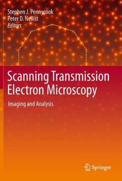
Scanning transmission electron microscopy has become a mainstream technique for imaging and analysis at atomic resolution and sensitivity, and the editors of this book are widely credited with bringing the field to its present popularity. Scanning Transmission Electron Microscopy: Imaging and Analysis provides a comprehensive explanation of the theory and practice of STEM from introductory to advanced levels, covering the history of the field, the instrument, image formation and scattering theory, as well as practical aspects of imaging and analysis. The authors present examples of the use of…mehr

  • Geräte: PC
  • ohne Kopierschutz
  • eBook Hilfe
  • Größe: 38.16MB
Produktbeschreibung
Scanning transmission electron microscopy has become a mainstream technique for imaging and analysis at atomic resolution and sensitivity, and the editors of this book are widely credited with bringing the field to its present popularity. Scanning Transmission Electron Microscopy: Imaging and Analysis provides a comprehensive explanation of the theory and practice of STEM from introductory to advanced levels, covering the history of the field, the instrument, image formation and scattering theory, as well as practical aspects of imaging and analysis. The authors present examples of the use of combined imaging and spectroscopy for solving materials problems in a variety of fields, including condensed matter physics, materials science, catalysis, and nanoscience. Therefore this is a comprehensive reference for those working in applied fields wishing to use the technique, for graduate students learning microscopy for the first time, and for specialists in other fields of microscopy.

Dieser Download kann aus rechtlichen Gründen nur mit Rechnungsadresse in A, B, BG, CY, CZ, D, DK, EW, E, FIN, F, GR, HR, H, IRL, I, LT, L, LR, M, NL, PL, P, R, S, SLO, SK ausgeliefert werden.

Autorenporträt
Stephen J. Pennycook obtained his B.A. degree in natural sciences from the University of Cambridge, England, in 1975, and his M.A. and Ph.D. degrees in physics from the same institution in 1978. He then continued at the University of Cambridge Cavendish Laboratory in postdoctoral positions until moving to the ORNL Solid State Division in 1982, where he is now the leader of the Electron Microscopy Group. His main research interest is the study of materials through the technique of Z-contrast scanning transmission electron microscopy (STEM). This technique provides a directly interpretable image of materials at the atomic scale, in which higher atomic numbers (Z) show brighter. It overcomes the phase problem associated with conventional electron microscopy and diffraction techniques by establishing incoherent imaging conditions, the electron equivalent of incoherent imaging in the optical microscope first described by Lord Rayleigh in 1895. Currently, the Solid State Division's 300-kV STEM produces the world's smallest electron probe, just 1.3 angstroms in diameter. The development of the Z-contrast technique has earned Dr. Pennycook an R&D 100 Award, the Heinrich Award from the Microbeam Analysis Society, a U.S. Department of Energy (DOE) Award for Outstanding Achievement in Solid State Sciences, and the Materials Research Society Medal. Recently, his research has focused on grain boundaries in ceramics, which resulted in a DOE Award for Outstanding Achievement in Metallurgy and Ceramics, as well as applications to optoelectronic materials, catalysts, and nanoparticles. Dr Peter D. Nellist's research centres on the applications and development of high-resolution electron microscope techniques, in particular scanning transmission electron microscopy (STEM), including atomic resolution Z-contrast imaging, electron energy-loss spectroscopy and applications of spherical aberration correctors. His technique development work includes methods for the three-dimensional imaging and spectroscopy of materials, and methods to allow high resolution imaging and spectroscopy of radiation sensitive materials. The aim is to use microscopy data in a quantitative way to make measurements of the atomic and electronic structure of materials.
Rezensionen
From the reviews:

"To describe in 18 chapters the current status in a wide field, a dazzling list of no less than 44 distinguished authors has been assembled. Fortunately, the role of the editors has continued well beyond the point of producing their own chapters to ensure that these different contributions are reasonably well integrated with a useful index....The editors' assertion that the experiment of focusing a beam of electrons down to an atomic scale and measuring its scattering has spectacular outcomes is most abundantly proved here."

--Archie Howie, Microscopy and Microanalysis

"The book opens with a magnificent 90-page history of STEM by S.J. Pennycook, which traces the story of the instrument from von Ardenne's microscope of the late 1930s to such very recent innovations as the confocal mode of operation. ... Very readable, lavishly illustrated and extremely thorough, this will remain a key publication on the history of the STEM." (Ultramicroscopy, Vol. 116, 2012)