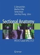
This timely atlas details advancements in PET/CT and SPECT/CT. Each chapter provides nuclear medicine practitioners, radiologists, oncologists, and residents with detailed information on normal anatomy of FDG PET/CT, variations and artifacts of FDG PET/CT, normal anatomy of non-FDG PET/CT, and normal anatomy of PET/CT and SPECT/CT. Coverage emphasizes anatomy to reinforce the names of organs and to support familiarization with normal and abnormal findings. The atlas has been compiled with help from experienced contributors from several top international imaging centers. Throughout the text, four-color images aid readers in proper interpretation.
Dieser Download kann aus rechtlichen Gründen nur mit Rechnungsadresse in A, B, BG, CY, CZ, D, DK, EW, E, FIN, F, GR, HR, H, IRL, I, LT, L, LR, M, NL, PL, P, R, S, SLO, SK ausgeliefert werden.
From the reviews:
"This book is an image-based guide to the sectional anatomy of fusion images obtained with PET/CT and SPECT/CT scanners. ... The book is aimed primarily at clinicians who routinely interpret images and is a useful reference for most molecular imaging probes currently used with PET/CT and SPECT/CT. ... this book would be a valuable resource for anyone reading PET/CT and SPECT/CT images, providing nuclear medicine and radiology physicians, especially with a practical reference for image interpretation." (Martin Allen-Auerbach, The Journal of Nuclear Medicine, Vol. 49, 2008)