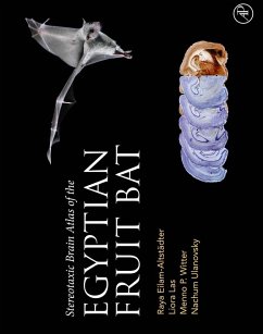
The Stereotaxic Brain Atlas of the Egyptian Fruit Bat provides the first stereotaxic atlas of the brain of the Egyptian fruit bat (
Rousettus aegyptiacus), an emerging model in neuroscience. This atlas contains coronal brain sections stained with cresyl violet (Nissl), AChE, and Parvalbumin - all stereotaxically calibrated. It will serve the needs of any neuroscientist who wishes to work with these bats - allowing to precisely target specific brain areas for electrophysiology, optogenetics, pharmacology, and lesioning. More broadly, this atlas will be useful to all neuroscientists working with bats, as it delineates many brain regions that were not delineated so far in any bat species. Finally, this atlas will provide a useful resource for researchers interested in comparative neuroanatomy of the mammalian brain.
- Provides detailed and accurate stereotaxic coverage of the Egyptian fruit bat forebrain
- Contains 87 plates of coronal sections of adult Egyptian fruit bats, each with one Nissl-stained hemisphere and the other stained either for AChE or Parvalbumin
- Delineates brain structures in the bat brain
- Serves as an essential tool for directing electrophysiology, imaging, optogenetics, pharmacology and lesioning in Egyptian fruit bats, and bats more generally
- Provides a rich resource for comparative neuroanatomy of the mammalian brain
Dieser Download kann aus rechtlichen Gründen nur mit Rechnungsadresse in A, B, BG, CY, CZ, D, DK, EW, E, FIN, F, GR, HR, H, IRL, I, LT, L, LR, M, NL, PL, P, R, S, SLO, SK ausgeliefert werden.

