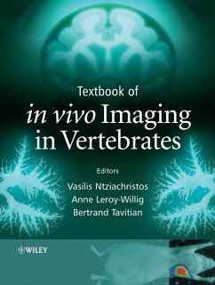
Textbook of in vivo Imaging in Vertebrates (eBook, PDF)
Redaktion: Ntziachristos, Vasilis; Tavitian, Bertrand; Leroy-Willig, Anne


Alle Infos zum eBook verschenken

Textbook of in vivo Imaging in Vertebrates (eBook, PDF)
Redaktion: Ntziachristos, Vasilis; Tavitian, Bertrand; Leroy-Willig, Anne
- Format: PDF
- Merkliste
- Auf die Merkliste
- Bewerten Bewerten
- Teilen
- Produkt teilen
- Produkterinnerung
- Produkterinnerung

Hier können Sie sich einloggen

Bitte loggen Sie sich zunächst in Ihr Kundenkonto ein oder registrieren Sie sich bei bücher.de, um das eBook-Abo tolino select nutzen zu können.
This book describes the new imaging techniques being developed to monitor physiological, cellular and subcellular function within living animals. This exciting field of imaging science brings together physics, chemistry, engineering, biology and medicine to yield powerful and versatile imaging approaches. By combining advanced non-invasive imaging technologies with new mechanisms for visualizing biochemical events and protein and gene function, non-invasive vertebrate imaging enables the in vivo study of biology and offers rapid routes from basic discovery to drug development and clinical…mehr
- Geräte: PC
- mit Kopierschutz
- eBook Hilfe
- Größe: 8.45MB
![Biomedical Sciences (eBook, PDF) Biomedical Sciences (eBook, PDF)]() Biomedical Sciences (eBook, PDF)82,99 €
Biomedical Sciences (eBook, PDF)82,99 €![Airway Smooth Muscle in Asthma and COPD (eBook, PDF) Airway Smooth Muscle in Asthma and COPD (eBook, PDF)]() Airway Smooth Muscle in Asthma and COPD (eBook, PDF)161,99 €
Airway Smooth Muscle in Asthma and COPD (eBook, PDF)161,99 €![An Introduction to Biomedical Science in Professional and Clinical Practice (eBook, PDF) An Introduction to Biomedical Science in Professional and Clinical Practice (eBook, PDF)]() Sarah J. PittAn Introduction to Biomedical Science in Professional and Clinical Practice (eBook, PDF)44,99 €
Sarah J. PittAn Introduction to Biomedical Science in Professional and Clinical Practice (eBook, PDF)44,99 €![Neurobiology of Motor Control (eBook, PDF) Neurobiology of Motor Control (eBook, PDF)]() Neurobiology of Motor Control (eBook, PDF)176,99 €
Neurobiology of Motor Control (eBook, PDF)176,99 €![Orientation and Navigation in Vertebrates (eBook, PDF) Orientation and Navigation in Vertebrates (eBook, PDF)]() Andrii RozhokOrientation and Navigation in Vertebrates (eBook, PDF)113,95 €
Andrii RozhokOrientation and Navigation in Vertebrates (eBook, PDF)113,95 €![Comparative Vertebrate Neuroanatomy (eBook, PDF) Comparative Vertebrate Neuroanatomy (eBook, PDF)]() Ann B. ButlerComparative Vertebrate Neuroanatomy (eBook, PDF)194,99 €
Ann B. ButlerComparative Vertebrate Neuroanatomy (eBook, PDF)194,99 €![Atlas of Developmental Field Anomalies of the Human Skeleton (eBook, PDF) Atlas of Developmental Field Anomalies of the Human Skeleton (eBook, PDF)]() Ethne BarnesAtlas of Developmental Field Anomalies of the Human Skeleton (eBook, PDF)144,99 €
Ethne BarnesAtlas of Developmental Field Anomalies of the Human Skeleton (eBook, PDF)144,99 €-
-
-
Dieser Download kann aus rechtlichen Gründen nur mit Rechnungsadresse in A, B, BG, CY, CZ, D, DK, EW, E, FIN, F, GR, HR, H, IRL, I, LT, L, LR, M, NL, PL, P, R, S, SLO, SK ausgeliefert werden.
- Produktdetails
- Verlag: John Wiley & Sons
- Seitenzahl: 388
- Erscheinungstermin: 20. August 2007
- Englisch
- ISBN-13: 9780470029589
- Artikelnr.: 37289883
- Verlag: John Wiley & Sons
- Seitenzahl: 388
- Erscheinungstermin: 20. August 2007
- Englisch
- ISBN-13: 9780470029589
- Artikelnr.: 37289883
- Herstellerkennzeichnung Die Herstellerinformationen sind derzeit nicht verfügbar.
). 4.0 Introduction. 4.1 Radioactivity. 4.2 Interaction of gamma rays with matter. 4.3 Radiotracer imaging with gamma emitters. 4.4 Detection of positron emitters. 4.5 Image properties and analysis. 4.6 Radiochemistry of gamma-emitting radiotracers. 4.7 Radiochemistry of positron-emitting radiotracers. 4.8 Major radiotracers and imaging applications. 5 Optical Imaging and Tomography (Antoine Soubret and Vasilis Ntziachristos). 5.0 Introduction. 5.1 Light - tissue interactions. 5.2 Light propagation in tissues. 5.3 Reconstruction and inverse problem. 5.4 Fluorescence molecular tomography (FMT). 6 Optical Microscopy in Small Animal Research (Rakesh K. Jain, Dai Fukumura, Lance Munn and Edward Brown). 6.0 Introduction. 6.1 Confocal laser scanning microscopy. 6.2 Multiphoton laser scanning microscopy. 6.3 Variants for In vivo imaging. 6.4 Surgical preparations. 6.5 Applications. 7 New Radiotracers, Reporter Probes and Contrast Agents (Coordinated by Bertrand Tavitian). 7.0 Introduction (Bertrand Tavitian). 7.1 New radiotracers (Bertrand Tavitian, Roberto Pasqualini and Frederic Dolle
). 7.2 Multimodal constructs for magnetic resonance imaging (Willem J.M. Mulder, Gustav J. Strijkers and Klaas Nicolay). 7.3 Fluorescence reporters for biomedical imaging (Benedict Law and Ching-Hsuan Tung). 7.4 New contrast agents for NMR (Silvio Aime). 7.5 Imaging techniques - reporter gene imaging agents (Huongfeng Li and Andreas H. Jacobs). 8 Multi-Modality Imaging (Coordinated by Vasilis Ntziachristos). 8.0 Introduction (Vasilis Ntziachristos). 8.1 Concurrent imaging versus computer-assisted registration (Fred S. Azar). 8.2 Combination of SPECT and CT (Jan Grimm). 8.3 FMT registration with MRI (Vasilis Ntziachristos). 9 Brain Imaging (Coordinated by Anne Leroy-Willig). 9.0 Introduction (Anne Leroy-Willig). 9.1 Bringing amyloid into focus with MRI microscopy (Greet Vanhoutte and Annemie Van der Linden). 9.2 Cerebral blood volume and BOLD contrast MRI unravels brain responses to ambient temperature fluctuations in fish (Annemie Van der Linden). 9.3 Assessment of functional and neuroanatomical re-organization after experimental stroke using MRI (Jet P. van der Zijden and Rick M. Dijkhuizen). 9.4 Brain activation and blood flow studies with speckle imaging (Andrew K. Dunn). 9.5 Manganese-enhanced MRI of the songbird brain: a dynamic window on rewiring brain circuits encoding a versatile behaviour (Vincent Van Meir and Annemie Van der Linden). 9.6 Functional MRI in awake behaving monkeys (Wim Vanduffel, Koen Nelissen, Denis Fize and Guy A. Orban). 9.7 Multimodal evaluation of mitochondrial impairment in a primate model of Huntington's disease (Vincent Lebon and Philippe Hantraye). 10 Imaging of Heart, Muscle, Vessels (Coordinated by Yves Fromes). 10.0 Introduction (Yves Fromes). 10.1 Cardiac structure and function (Yves Fromes). 10.2 Evaluation of therapeutic approaches in muscular dystrophy using MRI (Valerie Allamand). 10.3 Canine muscle oxygen saturation: evaluation and treatment of M-type phosphofructokinase deficiency (Kevin McCully and Urs Giger). 10.4 In vivo assessment of myocardial perfusion by NMR technology (Jorg. U.G. Streif, Matthias Nahrendorf and Wolfgang R. Bauer). 10.5 Ultrasound microimaging of strain in the mouse heart (F. Stuart Foster). 10.6 MR imaging of experimental atherosclerosis (Willem J.M. Mulder, Gustav J. Strijkers, Zahi A. Fayad and Klaas Nicolay). 11 Tumor Imaging (Coordinated by Vasilis Ntziachristos). 11.0 Introduction (Vasilis Ntziachristos). 11.1 Dynamic contrast-enhanced MRI of tumour angiogenesis (Charles Andre
Cuenod, Laure Fournier, Daniel Balvay, Clement Pradel, Nathalie Siauve and Olivier Clement). 11.2 Liver tumours: Evaluation by functional computed tomography (Charles Andre Cuenod, Laure Fournier, Nathalie Siauve and Olivier Clement). 11.3 Early detection of grafted Wilms' tumours (Erwan Jouannot). 11.4 Angiogenesis study using ultrasound imaging (Olivier Lucidarme). 11.5 Nuclear imaging of apoptosis in animal tumour models (Silvana Del Vecchio and Marco Salvatore). 11.6 Optical imaging of tumour-associated protease activity (Benedict Law and Ching-Hsuan Tung). 11.7 Tumour angiogenesis and blood flow (Rakesh K. Jain, Dai Fukumura, Lance L. Munn and Edward B. Brown). 11.8 Optical imaging of apoptosis in small animals (Eyk Schellenberger). 11.9 Fluorescence molecular tomography (FMT) of angiogenesis (Xavier Montet, Vasilis Ntziachristos, and Ralph Weissleder). 11.10 High resolution X-ray microtomography as a tool for imaging lung tumours in living mice (Nora De Clerck and Andrei Postnov). 12 Other Organs (Coordinated by Anne Leroy-Willig). 12.0 Introduction (Anne Leroy-Willig). 12.1 3D imaging of embryos and mouse organs by Optical Projection Tomography (James Sharpe). 12.2 Visualizing early Xenopus development with time lapse microscopic MRI (Cyrus Papan and Russell E. Jacobs). 12.3 Ultrasonic quantification of red blood cells development in mice (Johann Le Floch). 12.4 Placental perfusion MR imaging with contrast agent in a mouse model (Nathalie Siauve, Laurent Salomon and Charles Andre Cuenod). 12.5 Characterization of nephropathies and monitoring of renal stem cell therapies (Nicolas Grenier, Olivier Hauger, Yahsou Delmas and Christian Combe). 12.6 Optical imaging of lung inflammation (Jodi Haller). 12.7 Optical imaging in rheumatoid arthritis (Andreas Wunder). 13 Gene Therapy (Markus Klein and Andreas H. Jacobs). 13.0 Introduction. 13.1 Expression systems for genes of interest (GOI). 13.2 Gene delivery systems (vectors). 13.3 Suicide gene therapy. 13.4 Non-suicide gene therapy. 13.5 Imaging of gene expression. 13.6 Diseases targeted by gene therapy. 14 Cellular Therapies and Cell Tracking (Coordinated by Yves Fromes). 14.0 Introduction (Yves Fromes). 14.1 Are stem cells attracted by pathology? The case for cellular tracking by serial in vivo MRI (Michel Modo). 14.2 Cell tracking using MRI (V
t Herynek). 14.3 Cell labelling strategies for in vivo molecular MR imaging (Mathias Hoehn). 14.4 Animal imaging and medical challenges - cell labelling and molecular imaging (Yannic Waerzeggers, and Andreas H. Jacobs). Index.
