Thomas A. Woolsey, Joseph Hanaway, Mokhtar H. Gado
The Brain Atlas (eBook, ePUB)
A Visual Guide to the Human Central Nervous System
53,99 €
53,99 €
inkl. MwSt.
Sofort per Download lieferbar
0 °P sammeln
53,99 €
Als Download kaufen
53,99 €
inkl. MwSt.
Sofort per Download lieferbar
0 °P sammeln
Jetzt verschenken
Alle Infos zum eBook verschenken
53,99 €
inkl. MwSt.
Sofort per Download lieferbar
Alle Infos zum eBook verschenken
0 °P sammeln
Thomas A. Woolsey, Joseph Hanaway, Mokhtar H. Gado
The Brain Atlas (eBook, ePUB)
A Visual Guide to the Human Central Nervous System
- Format: ePub
- Merkliste
- Auf die Merkliste
- Bewerten Bewerten
- Teilen
- Produkt teilen
- Produkterinnerung
- Produkterinnerung
Bitte loggen Sie sich zunächst in Ihr Kundenkonto ein oder registrieren Sie sich bei
bücher.de, um das eBook-Abo tolino select nutzen zu können.
Hier können Sie sich einloggen
Sie sind bereits eingeloggt. Klicken Sie auf 2. tolino select Abo, um fortzufahren.
Bitte loggen Sie sich zunächst in Ihr Kundenkonto ein oder registrieren Sie sich bei bücher.de, um das eBook-Abo tolino select nutzen zu können.
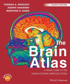
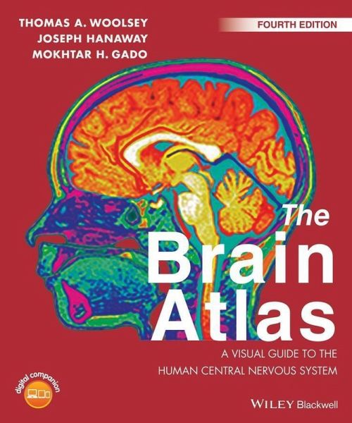
The Brain Atlas: A Visual Guide to the Human Central Nervous System integrates modern neuroscience with clinical practice and is now significantly revised and updated for a Fourth Edition. The book's five sections cover: Background Information, The Brain and Its Blood Vessels, Brain Slices, Histological Sections, and Pathways. These are depicted in over 350 high quality intricate figures making it the best available visual guide to human neuroanatomy.
- Geräte: eReader
- ohne Kopierschutz
- eBook Hilfe
Andere Kunden interessierten sich auch für
- Thomas A. WoolseyThe Brain Atlas (eBook, PDF)53,99 €
- Thomas A. WoolseyThe Brain Atlas (eBook, ePUB)65,99 €
- Neuropsychology for Occupational Therapists (eBook, ePUB)34,99 €
- Obsessive Compulsive Disorder (eBook, ePUB)80,99 €
- Oliver SacksThe Man Who Mistook His Wife for a Hat (eBook, ePUB)11,95 €
- The Treatment of Epilepsy (eBook, ePUB)278,99 €
- Roger A. BarkerNeuroanatomy and Neuroscience at a Glance (eBook, ePUB)28,99 €
-
-
-
The Brain Atlas: A Visual Guide to the Human Central Nervous System integrates modern neuroscience with clinical practice and is now significantly revised and updated for a Fourth Edition. The book's five sections cover: Background Information, The Brain and Its Blood Vessels, Brain Slices, Histological Sections, and Pathways. These are depicted in over 350 high quality intricate figures making it the best available visual guide to human neuroanatomy.
Dieser Download kann aus rechtlichen Gründen nur mit Rechnungsadresse in D ausgeliefert werden.
Produktdetails
- Produktdetails
- Verlag: Jossey-Bass
- Erscheinungstermin: 19. Januar 2017
- Englisch
- ISBN-13: 9781118438763
- Artikelnr.: 47516095
- Verlag: Jossey-Bass
- Erscheinungstermin: 19. Januar 2017
- Englisch
- ISBN-13: 9781118438763
- Artikelnr.: 47516095
- Herstellerkennzeichnung Die Herstellerinformationen sind derzeit nicht verfügbar.
Thomas A. Woolsey MD, lecturer in Biology; George H. and Ethel R. Bishop Scholar in Neuroscience Emeritus; Professor Emeritus of Experimental Neurological Surgery, of Experimental Neurology, of Anatomy and Neurobiology, of Cell Biology and Physiology and of Biomedical Engineering, Washington University School of Medicine, St Louis, MO, USA.
Dr Woolsey a world-renowned neurobiologist best known for his discovery of the cortical barrels in rodents. During his nearly forty years at Washington University School of Medicine, he has won numerous awards for teaching medical students, residents, graduate students, and undergraduates in psychology and life sciences.
Joseph Hanaway, MD, retired Clinical Assistant Professor of Neurology, Washington University School of Medicine, St Louis, MO, USA.
Dr Hanway has practiced neurology for over thirty years and has taught basic and clinical neuroanatomy at Harvard University, the University of Virginia, Washington University, and the University of Missouri Medical School.
Mokhtar H. Gado, MD, formerly Professor of Radiology, Mallinckrodt Institute of Radiology, Washington University School of Medicine, St Louis, MO, USA.
Dr Gado was an internationally known neuroradiologist with more than thirty-five years of clinical, research, and teaching experience at Washington University School of Medicine and the Mallinckrodt Institute of Radiology.
Preface xi
Acknowledgments xii
About the Digital Companion xiii
Part I Introduction
Overview 4
The Nervous System 5
Cells 5
Gray Matter/White Matter 5
Connections 7
CNS/PNS 7
Constitution of the CNS 7
Principal Divisions of the Brain 8
Cranial Nerves and Spinal Segments 8
Cerebrospinal Fluid and Its Circulation 9
Cortical Areas 10
Using this Book 10
Terminology 10
Terms of Relation-The Special Case of the Human Head 12
Labels 12
Image Groups 13
Pathways 14
Navigation 14
Materials and Methods 14
Subjects 14
Brain Specimens 15
Radiological Imaging 16
References 17
Part II The CNS and Its Blood VesselsnBrain 20
Cerebral Hemisphere and Brain Stem; Sulci and Gyri-Lateral Aspect 20
Cerebral Hemisphere and Brain Stem Arteries; Arteries of the Insula and
Lateral Sulcus; Arterial Territories-Lateral Aspect 22
Cerebral Hemisphere and Brain Stem; Sulci and Gyri-Mesial Aspect 24
Cerebral Hemisphere and Brain Stem Arteries; Arterial Territories-Mesial
Aspect 26
Cerebral Hemisphere and Brain Stem Arteries; by Conventional Angiography;
by MRA-Lateral Projection 28
Dural Venous Sinuses and Folds (Diagrammatic); by Conventional Angiography;
by MRV-Lateral Projection 30
Cerebral Hemispheres, Brain Stem, and Arteries; by MRA-Anterior Aspect 32
Cerebral Hemisphere and Brain Stem Arteries and Veins by Conventional
Angiography and Veins by MRV-Posteroanterior Projections 34
Cerebral Hemispheres and Brain Stem; Sulci and Gyri--Inferior Aspect 36
Cerebral Hemispheres and Brain Stem: Arteries and Cranial Nerves; Arterial
Territories; Axial MRA-Inferior Aspect 38
Brain Stem 40
Brain Stem, Diencephalon, Basal Ganglia, and Cerebellum-Anterolateral
Aspect 40
Brain Stem, Diencephalon, Basal Ganglia, and Cerebellum; Arteries and
Cranial Nerves-Anterolateral Aspect 41
Brain Stem, Diencephalon, Basal Ganglia, and Cerebellum; Arterial
Territories - Anterolateral Aspect 42
Brain Stem, Thalamus, and Striatum- Anterior Aspect 43
Brain Stem, Thalamus, and Striatum- Posterior Aspect 44
Brain Stem, Thalamus, and Striatum- Lateral Aspect 45
Cerebellum 46
Cerebellum-Superior Surface 46
Cerebellum-Inferior Surface 47
Spinal Cord 48
Arteries to Spinal Cord (Diagrammatic) 48
Segmental Arterial Supply of Spinal Cord (Diagrammatic) 49
Fiber Bundles 50
Principal Fiber Bundles in Cerebral Hemisphere and Brain Stem
(Semi-Schematic)-Lateral and Mesial Aspects 50
Principal Fiber Bundles in Coronal, Axial, and Sagittal Brain Sections
(Semi-Schematic) 51
Part III Brain Slices Coronal Sections 54
Coronal Section Through Rostral Wall of Lateral Ventricle with Vessel
Territories 54
Coronal Section Through Anterior Limit of Putamen with MRI 56
Coronal Section Through Head of Caudate Nucleus and Putamen with MRI 58
Coronal Section Through Anterior Limit of Amygdala with Vessel Territories
60
Coronal Section Through Tuber Cinereum with MRI 62
Coronal Section Through Interventricular Foramen (Foramen of Monro) with
Vessel Territories 64
Coronal Section Through Anterior Nucleus of Thalamus with MRI 66
Coronal Section Through Mamillothalamic Tract (Fasciculus) with Vessel
Territories 68
Coronal Section Through Mamillary Bodies with MRI 70
Coronal Section Through Subthalamic Nucleus with Vessel Territories 72
Coronal Section Through Posterior Limit of Interpeduncular Fossa with MRI
74
Coronal Section Through Posterior Commissure with Vessel Territories 76
Coronal Section Through Commissure of Superior Colliculi with MRI 78
Coronal Section Through Quadrigeminal Plate with Vessel Territories 80
Coronal Section Through Fourth Ventricle (IV) with MRI 82
Coronal Section Through Posterior Limit of Hippocampus with Vessel
Territories 84
Coronal Section Through Posterior Horns of Lateral Ventricles with MRI 86
Sagittal Sections 88
Sagittal Section Through Superior, Middle, and Inferior Temporal Gyri with
Vessel Territories 88
Sagittal Section Through Insula with MRI 90
Sagittal Section Through Claustrum and Lateral Putamen with Vessel
Territories 92
Sagittal Section Through Lateral Putamen with MRI 94
Sagittal Section Through Termination of Optic Tract with MRI 96
Sagittal Section Through Pulvinar with Vessel Territories 98
Sagittal Section Through Ambient Cistern with MRI 100
Sagittal Section Through Olfactory Tract with Vessel Territories 102
Sagittal Section Through Inferior Cerebellar Peduncle (Restiform Body) with
Vessel Territories 104
Sagittal Section Through Superior Cerebellar Peduncle (Brachium
Conjunctivum) with MRI 106
Sagittal Section Through Red Nucleus with Vessel Territories 108
Sagittal Section Through Cerebral Aqueduct (Aqueduct of Sylvius) with MRI
110
Axial Sections 112
Axial Section Through Superior Caudate Nucleus with MRI 112
Axial Section Through Inferior Corpus Callosum with Vessel Territories 114
Axial Section Through Superior Putamen with MRI 116
Axial Section Through Putamen with Vessel Territories 118
Axial Section Through Frontoparietal Opercula with MRI 120
Axial Section Through Midlevel Diencephalon with Vessel Territories 122
Axial Section Through Anterior Commissure with MRI 124
Axial Section Through Habenular Commissure with Vessel Territories 126
Axial Section Through Superior Colliculi with MRI 128
Axial Section Through Anterior Perforated Substance with Vessel Territories
130
Axial Section Through Inferior Colliculi with MRI 132
Part IV Histological Sections Cerebellum 136
Horizontal Section Through Fastigial Nucleus with Vessel Territories 136
Horizontal Section Through Dentate Nucleus with MRI 137
Brain Stem 138
Transverse Section Through Superior Colliculus with Vessel Territories 138
Transverse Section Through Oculomotor Nucleus with MRI 139
Transverse Section Through Inferior Colliculus with Vessel Territories 140
Transverse Section Through Superior Pons and Isthmus with MRI 141
Transverse Section Through Middle Pons with Vessel Territories 142
Transverse Section Through Facial Genu with MRI 143
Transverse Section Through Vestibulocochlear Nerve Root with Vessel
Territories 144
Transverse Section Through Glossopharyngeal Nerve Root with MRI 145
Transverse Section Through Fourth Ventricle with Vessel Territories 146
Transverse Section Through Hypoglossal Nucleus with MRI 147
Transverse Section Through Inferior Olive with Vessel Territories 148
Transverse Section Through Decussation of Pyramids with MRI 149
Spinal Cord 150
Transverse Section Through Upper Cervical Level with Vessel Territories 150
Transverse Section Through Cervical Enlargement with MRI 151
Transverse Section Through Thoracic Level with Vessel Territories 152
Transverse Section Through Lumbar Enlargement with Vessel Territories 153
Transverse Section Through Sacral Level 154
Basal Ganglia and Thalamus 155
Coronal Section Through Nucleus Accumbens 155
Coronal Section Through Optic Chiasm 156
Coronal Section Through Anterior Commisure 157
Coronal Section Through Anterior Thalamic Tubercle 158
Coronal Section Through Mamillothalamic Tract 159
Coronal Section Through H Fields of Forel 160
Coronal Section Through Dorsal Lateral Geniculate Nucleus 161
Coronal Section Through Pulvinar 162
Hypothalamus 163
Coronal Section Through Optic Chiasm; Coronal Section Through Pituitary
Stalk 163
Coronal Section Through Interthalamic Adhesion; Coronal Section Through
Mamillary Bodies 164
Basal Forebrain 165
Coronal Section Through Olfactory Trigone and Nucleus Basalis 165
Hippocampus 166
Coronal Section Through Body of Hippocampus 166
Part V Pathways Brain Stem 170
General Organization of Spinal Cord Gray Matter 170
General Organization of Cranial Nerve Gray Matter 172
Sensory Cranial Nerves and Nuclei 174
Motor Cranial Nerves and Nuclei 175
Organization of Cranial Nerve Nuclei into Columns-Posterior Aspect 176
Organization of Cranial Nerve Nuclei into Columns-Anterior Aspect 177
Thalamus 178
Organization of the Thalamus 178
Hypothalamus 180
Organization of the Hypothalamus 180
Sensory Systems 182
Touch and Position Sense Pathways: Posterior (Dorsal) Column/Medial
Lemniscus and Trigeminal Main Sensory Nucleus 182
Touch Pathways: Anterior and Lateral Spinothalamic Tracts and Trigeminal
Spinal Nucleus 184
Pain Pathways 186
Touch Pathways: Head and Face 188
Taste Pathways 190
Visual Pathways 192
Olfactory Pathways 194
Auditory Pathways 196
Sensory/Motor Systems 198
Vestibular Pathways 198
Motor Systems 200
Corticospinal (Pyramidal) and Corticobulbar Pathways 200
Rubrospinal and Tectospinal Pathways 202
Reticulospinal Pathways 204
Cerebellum 206
Cerebellar Pathways: Somatic Afferents 206
Cerebellar Pathways: Afferents (Non-Somatic) 208
Cerebellar Pathways: Efferents 210
Basal Ganglia 212
Basal Ganglia Pathways 212
Hippocampus 214
Hippocampal Pathways: Afferents 214
Hippocampal Pathways: Efferents 216
Amygdala 218
Amygdalar Pathways: Afferents 218
Amygdalar Pathways: Efferents 220
Hypothalamus 222
Hypothalamic Pathways: Afferents 222
Hypothalamic Pathways: Efferents 224
Arousal and Sleep Pathways 226
Hunger and Feeding Pathways 228
Circumventricular Organs 230
Autonomic Systems 232
Autonomic Pathways: Afferents 232
Autonomic Pathways: Sympathetic Efferents 234
Autonomic Pathways: Parasympathetic Efferents 236
Modulatory Systems 238
Cholinergic and Dopaminergic Pathways 238
Noradrenergic and Serotonergic Pathways 240
Index 243