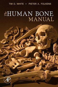
- Features more than 500 color photographs and illustrations in a portable format; most in 1:1 ratio
- Provides multiple views of every bone in the human body
- Includes tips on identifying any human bone or tooth
- Incorporates up-to-date references for further study
Dieser Download kann aus rechtlichen Gründen nur mit Rechnungsadresse in A, B, BG, CY, CZ, D, DK, EW, E, FIN, F, GR, HR, H, IRL, I, LT, L, LR, M, NL, PL, P, R, S, SLO, SK ausgeliefert werden.
"As a human osteology manual, this book is an excellent resource for archaeologists, palaeontologists and forensic anthropologists. It is a thoroughly written, well illustrated and can be highly recommended." --Francis Thackeray, Transvaal Museum, Pretoria, South Africa
"This volume is a compact form of previously highly successful editions of Human Osteology by these same authors. In its newest form it is somewhat abbreviated, but is nevertheless an extraordinarily accurate and completeguide to all of the fundamentals of osteoanthropology. The illustrations retain their same vivid three dimensional quality as in previous editions, and the accuracy and thorough review of all major methods (aging, sexing, excavation, laboratory care, etc.) have been retained. It is the perfect companion text and manual for any undergraduate or graduate human osteology course, and is equally highly recommended as well to students of medicine, dentistry, physical therapy, chiropractic, etc., simply because it presents such clear and instructive presentations of fundamental human anatomy. It is, quite simply, the best human anatomy manual currently available." --C. Owen Lovejoy, Kent State University

