129,95 €
129,95 €
inkl. MwSt.
Sofort per Download lieferbar
payback
65 °P sammeln
129,95 €
129,95 €
inkl. MwSt.
Sofort per Download lieferbar

Alle Infos zum eBook verschenken
payback
65 °P sammeln
Als Download kaufen
129,95 €
inkl. MwSt.
Sofort per Download lieferbar
payback
65 °P sammeln
Jetzt verschenken
129,95 €
inkl. MwSt.
Sofort per Download lieferbar

Alle Infos zum eBook verschenken
payback
65 °P sammeln
  • Format: PDF


This book covers the imaging of thoracic structures with a focus on evaluation of the lungs. Pulmonary conditions are the most common global cause of mortality and morbidity, with lung cancer and tuberculosis being prevalent diseases in the developed and developing world, respectively. In the last few years there have been rapid changes in the imaging interpretation of lung diseases, particularly imaging of idiopathic pulmonary fibrosis. However, updated literature is not available yet. Filling this gap, the book provides radiologists with up-to-date, handy information on reading chest…mehr

  • Geräte: PC
  • ohne Kopierschutz
  • eBook Hilfe
  • Größe: 82.92MB
Produktbeschreibung


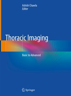
This book covers the imaging of thoracic structures with a focus on evaluation of the lungs. Pulmonary conditions are the most common global cause of mortality and morbidity, with lung cancer and tuberculosis being prevalent diseases in the developed and developing world, respectively. In the last few years there have been rapid changes in the imaging interpretation of lung diseases, particularly imaging of idiopathic pulmonary fibrosis. However, updated literature is not available yet. Filling this gap, the book provides radiologists with up-to-date, handy information on reading chest scans.

This book uses standard terms and lexicons to describe the imaging findings. It consists of 14 chapters, and includes more than 1200 illustrations covering both commonly and rarely encountered conditions. The first chapter discusses imaging patterns and signs in thoracic imaging, while the chapter on interstitial lung diseases describes the hallmarks that are useful in the diagnostic pathway of these diseases. This chapter also incorporates the latest diagnostic categories of UIP based on CT patterns, and provides pictorial examples. Written by experts in the field, the book highlights imaging features and relevant clinical findings that may help in reaching diagnosis, and wherever possible, it presents the imaging features in tables for ease of memorizing.


Dieser Download kann aus rechtlichen Gründen nur mit Rechnungsadresse in A, B, BG, CY, CZ, D, DK, EW, E, FIN, F, GR, HR, H, IRL, I, LT, L, LR, M, NL, PL, P, R, S, SLO, SK ausgeliefert werden.

Autorenporträt
Dr. Ashish Chawla is an American Board of Radiology certified radiologist. He completed his 2-year fellowship in cardiothoracic radiology at the University of Colorado Denver's School of Medicine and the National Jewish Health.  He is Head of the Cardiopulmonary Imaging section, Senior Consultant and Research Lead at the Department of Diagnostic Radiology in Khoo Teck Puat Hospital, Singapore. He is also an adviser to the CT Imaging section. His research interests include imaging of the chest in the emergency department and diffuse lung diseases. He has authored more than 60 peer-reviewed publications, presented numerous posters and delivered lectures at international conferences. He is actively involved in radiology education and regularly organizes thoracic radiology educational courses.