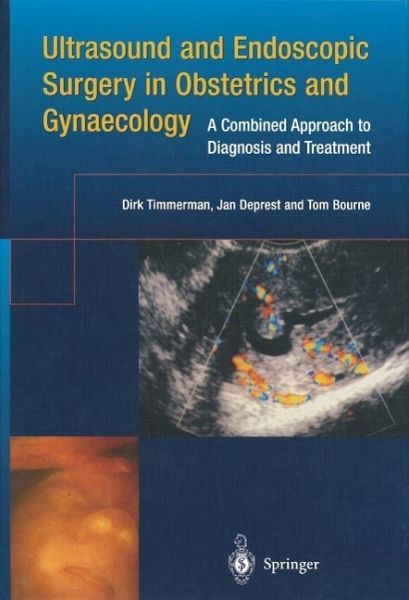
Ultrasound and Endoscopic Surgery in Obstetrics and Gynaecology (eBook, PDF)
A Combined Approach to Diagnosis and Treatment
Versandkostenfrei!
Sofort per Download lieferbar
40,95 €
inkl. MwSt.
Weitere Ausgaben:

PAYBACK Punkte
20 °P sammeln!
Unique in that it demonstrates the management of common gynecological conditions through accurate ultrasound diagnosis and minimally invasive treatment, this text highlights the pivotal role these two techniques play in modern obstetrics and gynecology. This authoritative text includes sections on menorrhagia, postmenopausal bleeding, endometrial malignancy, urogynecology, ovarian masses, endometriosis, early pregnancy complications and infertility. Multi-contributed, with short, practical chapters, the book is well illustrated with over 70 color illustrations.
Dieser Download kann aus rechtlichen Gründen nur mit Rechnungsadresse in A, B, BG, CY, CZ, D, DK, EW, E, FIN, F, GR, HR, H, IRL, I, LT, L, LR, M, NL, PL, P, R, S, SLO, SK ausgeliefert werden.


