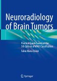
While several books describing imaging of brain tumors from MR acquisition techniques to differential diagnosis are written by different contributors and present chapters with different styles and design, this book illustrates a unique vision and structure putting together modern molecular classification of brain tumor with modern neuroradiology.
After an introduction on general imaging features of brain tumors the book explores each different tumor according to 2021 WHO classification, distinguishing however between adult and pediatric tumors, being the epidemiology substantially different between these two groups.
The approach is schematic with few essential information on epidemiology, genetics, clinical features, location and prognosis, followed by a detailed description of imaging features with a large number of examples. Figures are mainly put together with the same modality considering all the different MR techniques as well as CT when it can be useful. Each figure provides T1, T2, FLAIR, DWI, ADC, perfusion imaging techniques, spectroscopy and post contrast study. Some examples of Amide Proton Transfer (APT) technique are provided as well.
At the end of each chapter a scheme summarizes the different appearance of the tumor in any different sequence.
This book will be an invaluable tool for neuroradiologists, radiologists, neurosurgeons, neurologists, pediatricians, and pathologists.
Hinweis: Dieser Artikel kann nur an eine deutsche Lieferadresse ausgeliefert werden.


