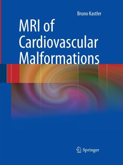
This book represents a new and fundamentally different way of learning cardiac magnetic resonance (CMR). Unlike previous CMR textbooks containing a few pre-selected views for each case, this book contains the entire scan for each of the 150 clinical cases (totalling more than 88,000 CMR images). Within each case, all the necessary tools to make the diagnosis are presented in exactly the same format as they are used every day at leading CMR clinical centers, including side-by-side movies, image magnification, single stepping, and even measurement tools (measurements require an internet connection). This format provides the student of CMR with the unprecedented opportunity to review all of the data and attempt to reach their own conclusion, and then reference an expert opinion simply by clicking on the "Case Discussion" web link.
The cases demonstrate the utility of CMR for the entire spectrum of cardiovascular pathology. Straightforward cases are presented, along with more unusual entities. The first few cases are designed to introduce the reader to the display format and to the techniques used in CMR, including the mechanics and nuances of image acquisition. The cases that then follow can be sorted by category (congenital heart disease, cardiomyopathies, etc) if desired, but are otherwise presented in a random format to allow readers to test themselves in interpreting "unknowns". In the initial presentation of the images, the history is withheld, but clicking the "history" button at the top of the page will reveal it. Through this presentation of unknown cases, the reader can encounter the unfiltered "raw data" of real patient scans, and can formulate their own opinion, then obtaining immediate feedback by reading the discussion.
The print part of this project is organized into two sections: one is an overview of CMR imaging with an introduction to current techniques, applications, and protocols and the second includes the Case Discussions that explain each case. The printed material is thus available for offline reading.
Cardiovascular MR imaging has become a robust, clinically useful mod- ity, and the rapid pace of innovation and important information it conveys have attracted many students whose goal is to become adept practitioners. In turn, many excellent textbooks have been written to aid this process. These books are necessary and useful in helping the student learn the underlying pulse sequences used in CMR, as well as the imaging findings in a variety of disorders. However, one of the difficulties inherent in learning CMR from a book is that the printed format is not the ideal medium to d- play the dynamic imaging that comprises a typical CMR case. For instance, it may be difficult to perceive focal areas of wall motion abnormality on serial static pictures, but these abnormalities are often easily seen on cine loops. One might say that trying to learn CMR solely from a standard textbook with illustrations is like trying to learn to drive by looking at snapshots obtained through the windshield of a moving car. The learner needs to see the cardiac motion and decide if it is normal or abnormal; he or she needs to be in the driver's seat. An additional limitation of the ava- able textbooks on CMR is that while they often have superb illustrations of abnormal findings, these images have been preselected.

