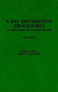
Harold P Klug, LeRoy E Alexander
X-Ray Diffraction Procedures
For Polycrystalline and Amorphous Materials
Harold P Klug, LeRoy E Alexander
X-Ray Diffraction Procedures
For Polycrystalline and Amorphous Materials
- Gebundenes Buch
- Merkliste
- Auf die Merkliste
- Bewerten Bewerten
- Teilen
- Produkt teilen
- Produkterinnerung
- Produkterinnerung
A complete view of x-ray diffraction procedures For those working in the field who wish to go beyond push-button applications, X-Ray Diffraction Procedures for Polycrystalline and Amorphous Materials provides a strong guide to the science and practical techniques of geometrical crystallography and x-ray diffraction of crystals. The book then moves on to provide more complete coverage of space lattices, point groups and space groups, the production of x-rays, measurement of x-rays, photographic powder techniques, and a host of other related topics. The book is then rounded out with a number of appendices for further reference.…mehr
Andere Kunden interessierten sich auch für
![Structure Determination from Powder Diffraction Data Structure Determination from Powder Diffraction Data]() W.I.F. David / K. Shankland / L.B. McCusker / Ch. Baerlocher (eds.)Structure Determination from Powder Diffraction Data319,99 €
W.I.F. David / K. Shankland / L.B. McCusker / Ch. Baerlocher (eds.)Structure Determination from Powder Diffraction Data319,99 €![International Tables for Crystallography, Volume F International Tables for Crystallography, Volume F]() International Tables for Crystallography, Volume F467,99 €
International Tables for Crystallography, Volume F467,99 €![Diffuse X-Ray Scattering and Models of Disorder Diffuse X-Ray Scattering and Models of Disorder]() T R WelberryDiffuse X-Ray Scattering and Models of Disorder143,99 €
T R WelberryDiffuse X-Ray Scattering and Models of Disorder143,99 €![Crystal Structure Analysis Crystal Structure Analysis]() Peter MainCrystal Structure Analysis206,99 €
Peter MainCrystal Structure Analysis206,99 €![X-Ray Diffraction by Disordered Lamellar Structures X-Ray Diffraction by Disordered Lamellar Structures]() Victor A. DritsX-Ray Diffraction by Disordered Lamellar Structures81,99 €
Victor A. DritsX-Ray Diffraction by Disordered Lamellar Structures81,99 €![Plasticity of Crystals With Special Reference to Metals Plasticity of Crystals With Special Reference to Metals]() Erich SchmidPlasticity of Crystals With Special Reference to Metals40,99 €
Erich SchmidPlasticity of Crystals With Special Reference to Metals40,99 €![Fast Reactions in Solids Fast Reactions in Solids]() Frank Philip BowdenFast Reactions in Solids35,99 €
Frank Philip BowdenFast Reactions in Solids35,99 €-
-
-
A complete view of x-ray diffraction procedures For those working in the field who wish to go beyond push-button applications, X-Ray Diffraction Procedures for Polycrystalline and Amorphous Materials provides a strong guide to the science and practical techniques of geometrical crystallography and x-ray diffraction of crystals. The book then moves on to provide more complete coverage of space lattices, point groups and space groups, the production of x-rays, measurement of x-rays, photographic powder techniques, and a host of other related topics. The book is then rounded out with a number of appendices for further reference.
Hinweis: Dieser Artikel kann nur an eine deutsche Lieferadresse ausgeliefert werden.
Hinweis: Dieser Artikel kann nur an eine deutsche Lieferadresse ausgeliefert werden.
Produktdetails
- Produktdetails
- Verlag: Wiley
- 2nd Revised edition
- Seitenzahl: 992
- Erscheinungstermin: 28. Mai 1974
- Englisch
- Abmessung: 231mm x 161mm x 58mm
- Gewicht: 1294g
- ISBN-13: 9780471493693
- ISBN-10: 0471493694
- Artikelnr.: 22206563
- Herstellerkennzeichnung
- Libri GmbH
- Europaallee 1
- 36244 Bad Hersfeld
- gpsr@libri.de
Harold P. Klug and Leroy E. Alexander are the authors of X-Ray Diffraction Procedures: For Polycrystalline and Amorphous Materials, 2nd Edition, published by Wiley.
1. Elementary Crystallography. 1-1 The Crystalline State. 1-1.1 Crystalline and Amorphous Solids. 1-1.2 Definition of a Crystal. 1-1.3 Characteristics of the Crystalline and Vitreous States. 1-2 Crystal Geometry. 1-2.1 External Form and Habit of Crystals. 1-2.2 Constancy of Interfacial Angles. 1-2.3 Symmetry Elements of Crystals. 1-2.4 Pseudosymmetry, 13. 1-2.5 Crystallographic Axes. 1-2.6 Axial Ratios. 1-2.7 The Six Crystal Symmetry Systems. 1-2.8 Miller Indices. 1-2.9 The Law of Rational Indices. 1-2.10 Crystal Forms. 1-2.11 Composite Crystals and Twinning. 1-2.12 Equation for the Plane (hkl). 1-2.13 Zones and Zone Relationships. 1-3 Space Lattices. 1-3.1 Historical Introduction. 1-3.2 Definition. 1-3.3 The Unit Ceil. 1-3.4 The 14 Bravais Lattices. 1-3.5 Some Crystallographic Implications of Space Lattices. 1-3.6 Distance between Neighboring Lattice Planes in the Series (hkl). 1-3.7 The Reciprocal Lattice. 1-4 Point Groups and Space Groups. 1-4.1 The Point Group or Crystal Symmetry Class. 1-4.2 The Space Group. General References. Specific References. 2. The Production and Properties of X-rays. 2-1 X-Ray Safety and Protection. 2-2 The Production of X-Rays. 2-2.1 The Origin of X-Rays. 2-2.2 X-Ray Tubes. A. Gas tubes. B. Hot-cathode tubes. C. Modern diffraction tube design. D. Cold-cathode diffraction tubes. E. High-intensity diffraction tubes. F. Microfocus diffraction tubes. 2-2.3 Power Equipment for the Production of X-rays. 2-2.4 Commercial X-ray Generators for Diffraction. 2-2.5 Isotopic X-ray Sources. 2-3 Properties of X-Rays and their Measurement. 2-3.1 The X-ray Spectrum of an Element. A. The continuous x-ray spectrum. B. The characteristic x-ray spectrum. 2-3.2 The Precise Determination of X-ray Wavelengths. 2-3.3 Absorption of X-rays. 2-3.4 Secondary Fluorescent and Scattered X-rays. 2-3.5 Refraction of X-rays. 2-3.6 Monochromatization of X-radiation. A. Single filter technique. B. Balanced-filter technique. C. Crystal monochromator techniques. D. Graphite monochromators. 2-3.7 The Photographic Effects of X-rays. General References. Specific References. 3. Fundamental Principles of X-ray Diffraction. 3-1 Kinematical and Dynamical Diffraction Theory. 3-2 The Geometry of Diffraction. 3-2.1 Scattering of X-rays by Electrons and Atoms. 3-2.2 Scattering by a Regularly Spaced Row of Atoms. 3-2.3 Conditions for Diffraction by a Linear Lattice of Atoms. 3-2.4 Diffraction by a Simple Cubic Lattice. 3-2.5 Proof that the "Diffracting Plane" is a Lattice Plane. 3-2.6 The Bragg Equation. 3-2.7 Derivation of the Bragg Equation from the "Reflection" Analogy. 3-2.8 The Geometrical Picture of Diffraction in Reciprocal Space. 3-3 The Intensity of Diffraction. 3-3.1 Perfect and Imperfect Crystals. 3-3.2 Primary and Secondary Extinction. 3-3.3 Relative and Absolute Intensities. 3-3.4 Factors Affecting the Diffraction Intensities. A. The polarization factor. B. The Lorentz and "velocity" factors. C. The temperature factor. D. The atomic scattering factor. E. The structure factor. F. The multiplicity factor. G. The absorption factor. 3-3.5 Expressions for the Relative Intensity of Diffraction by the Various Techniques. 3-3.6 Lattice-Centering and Space-Group Extinctions. General References. Specific References. 4. Photographic Powder Techniques. 4-1 The Debye-Scherrer Method. 4-1.1 Introduction. 4-1.2 Camera Design. A. General geometry. B. Details of camera construction. C. Camera support and alignment. 4-1.3 Preparation of the Powder. 4-1.4 Mounting the Powder. 4-1.5 Making the Exposure. 4-1.6 Processing the Film. 4-2 Parafocusing Methods. 4-3 Monochromatic-Pinhole Techniques. 4-3.1 Forward-Reflection Method. 4-3.2 Back-Reflection Method. 4-4 Microcameras and Microbeam Techniques. 4-5 High-Temperature Techniques. 4-6 Low-Temperature Techniques. 4-7 High-Pressure Techniques. General References. Specific References. Diffractometric Powder Technique. 5-1 Geometry of the Powder Diffractometer. 5-1.1 General Features. 5-1.2 Details of the Optical Arrangement. 5-1.3 The Seemann-Bohlin Diffractometer. 5-1.4 Alignment and Angular Calibration of the Diffractometer. A. Operations appropriately performed in advance by the manufacturer. B. Further internal alignment of the goniometer. C. Alignment of the goniometer with respect to the x-ray tube. D. Calibration of the O° 2
position. E. Calibration of angular registration between 0 and 180° 2
. 5-2 Profiles and positions of diffraction maxima. 5-2.1 Convolution Synthesis of Line Profiles. A. X-ray source, gI. B. Flat specimen surface, gII. C. Axial divergence, gIII. D. Specimen transparency, gIV. E. Receiving slit, gv. F. Comparison of calculated and experimental line profiles. 5-2.2 Displacement and Breadth of Diffraction Maxima. A. Line position. B. Line breadth. C. The practical determination of the centroid and variance. 5-2.3 Accurate Determination of Interplanar (d) Spacings. 5-2.4 "Routine" Determination of Interplanar (d) Spacings. 5-3 Electrical Characteristics of the Diffractometer. 5-3.1 General Arrangement of Components. 5-3.2 Radiation Detectors (Quantum Counters). A. Gas-ionization counters. B. Geiger-Müller counters. C. Proportional counters. D. Scintillation counters. E. Solid-state (energy-dispersive) detectors. 5-3.3 Nonlinearity of Detector Response. 5-3.4 Monochromatizing Techniques. A. Pulse-height discrimination and analysis. B. Ross balanced filters. C. Crystal monochromators. 5-4 Choice of Experimental Conditions and Procedures. 5-4.1 Statistical Accuracy of Counter Measurements. 5-4.2 The Specimen. A. Preparation of powders. B. Rotation of the specimen. C. Preferred orientation and the specimen mount. D. High-temperature techniques. E. Low-temperature techniques. F. Other special specimen techniques. 5-4.3 Transmission Techniques. 5-4.4 Continuous-Scan Techniques. 5-4.5 Step-Scan Techniques and Automation. General References. Specific References. 6. The Interpretation of Powder Diffraction Data. 6-1 The Viewing and Precision Measurement of Powder Photographs. 6-2 Determination of Interplanar (d) Spacings. 6-2.1 Debye-Scherrer Patterns, 424. 6-2.2 Monochromatic-Pinhole (Flat-Film) Patterns, 435. 6-3 Indexing Cubic Powder Patterns. 6-3.1 Reciprocal-Lattice Picture of Diffraction by a Cubic Powder. 6-3.2 Indexing a Cubic Pattern by sin2
Ratios. 6-3.3 Determination of the Unit-Cell Dimension a. 6-3.4 Indexing a Cubic Pattern When a Is Known. 6-4 Determination of Lattice Type. 6-5 Indexing Noncubic Powder Patterns. 6-5.1 Indexing Noncubic Patterns When the Unit-Cell Dimensions are Known. 6-5.2 Graphical Methods of Indexing. 6-5.3 Analytical Methods of Indexing: Tetragonal, Hexagonal, and Orthorhombic Patterns. 6-5.4 Analytical Methods of Indexing: Monoclinic and Triclinic Patterns. 6-6 Automated Computing Procedures for Indexing Powder Patterns. 6-6.1 Programs for Patterns of Orthorhombic and Higher Symmetry. 6-6.2 Programs for Patterns of Low Symmetry. 6-7 The Measurement of Intensities from Photographic Blackening. 6-7.1 Introduction. 6-7.2 Preparation of a Graded Intensity Scale. 6-7.3 Visual Estimation of Intensities. 6-7.4 Photometer Techniques. 6-8 The Measurement of Intensities with the X-Ray Diffractometer. 6-9 Putting Intensities on an Absolute Scale. 6-10 Special Scattering and Diffraction Effects. 6-10.1 Background Effects. A. Background due to lattice imperfections. B. Background due to general radiation. C. Absorption discontinuities. D. Air scatter. E. Secondary fluorescence radiation. 6-10.2 Reflections of Unusual Character. A. Spotty lines. B. Arclike lines. C. Broadened lines. D. Two-dimensional lattice lines. E. Splitting of lines. 6-10.3 Spurions lines. A. Lines due to misalignment of camera elements. B. Diffraction effects from the sample mount. C. Diffraction from radiation contaminants. General References. Specific References. 7. Qualitative and Quantitative Analysis of Crystalline Powders. 7-1 Routine Qualitative Identification of Crystalline Powders. 7-1.1 The JCPDS Powder Diffraction File (PDF). 7-1.2 Experimental Technique of the PDF Method. A. Preparation of the diffraction pattern. B. Measurement of lines on films and diffractometer charts. C. Identification interpretation of the data. 7-1.3 Computer Applications in the PDF Method. 7-1.4 Complications and Limitations of the PDF Method. 7-1.5 Special Identification Techniques. A. Compound identification by isomorphism. B. Procedures for or ganic compounds. C. Identification of clay minerals. 7-2 Quantitative Analysis of Powder Mixtures. 7-2.1 Basic Aspects of Absorption in Quantitative Analysis. A. Mixtures of N components: µ*J = ???*. B. Mixtures of two components: µ*1 ??? µ*M. > 2); µ*1 ??? µ*M. 7-2.2 Photographic-Microphotometric Technique. 7-2.3 Counter Diffractometric Technique. A. Instrumental requirements. B. General recommendations on procedure. 7-2.4 Outline of Important Analytical Procedures. A. Direct analysis when µ*J = ???*. B. Direct analysis of two-component syatems, µ*1 ??? µ*2. C. Direct analysis by absorption-diffraction, multicomponent systems. D. Internal-standard analysis for one component of a multicomponent system, no interfering lines. E. Internal-standard analysis for one component, interfering lines of unknown and standard. F. Simultaneous analysis for several components with allowance for line superpositions. G. Analysis by dilution of sample with yJ grams of unknown per gram of sample. 7-2.5 Selected Examples and Applications. A. Dust analysis. B. Retained austenite in steel. C. Organic mixtures. D. Miscellaneous inorganic analyses. E. Analysis of solid-solution phases. General References. Specific References. 8. The Precision Determination of Lattice Constants. 8-l General Considerations. 8-2 Sources of Systematic Errors in the Debye-Scherrer Method. 8-2.1 Radius Errors and Film Shrinkage. 8-2.2 Specimen Eccentricity. 8-2.3 Sample Absorption and Radial Divergence of the Beam. A. Bradley and Jay's approximate treatment. B. More rigorous investigations of the absorption error. 8-2.4 Axial Divergence of the Beam. 8-3 Methods of Correcting for Errors in the Debye-Scherrer Method. 8-3.1 Use of Calibrating Substances. 8-3.2 The Straumanis Method of Refined Experimental Technique. A. Essential features of the Straumanis method. B. Illustrative film measurements and calculations. 8-3.3 The Convolution-Film Method with the Likelihood Ratio Method. A. The Convolution-Film Method (CFM). B. The Likelihood Ratio Method (LRM). C. Application of the Convolution-Film Method to IUCr Silicon. 8-3.4 Use of Extrapolation Methods. A. Bradley and Jay's extrapolation against cos2
. B. Extrapolation against (cos2
)/sin
+ (cos2
)/
. C. Cohen's Least-Squares Extrapolation. 8-4 Precise Lattice Constants by other Film Techniques. 8-5 Precise Lattice Constants from Diffractometric Measurements. 8-6 The Precision Determination of Lattice Constants of Noncubic Materials. 8-7 Summary. General References. Specific References. 9. Crystallite Size and Lattice Strains From Line Broadening. 9-1 Determination of the Pure Line Profile. 9-1.1 The Fourier-Transform Method. The Rachinger Correction. 9-1.2 The Method of Iterative Folding. 9-1.3 Simplified Methods. Gaussian or Cauchy Profiles. B. Jones' Correction Curves for Debye-Scherrer Lines. C. The K
1
2 Doublet Correction for the Debye-Scherrer Technique. D. Correction Curves for Diffractometer Line Profiles. 9-2 Determination of Crystallite Size and Lattice Imperfections Simultaneously. 9-2.1 Introduction. 9-2.2 The Fourier Method of Warren and Averbach. A. Derivation of the Fourier series expression. B. Separation of size and distortion components. C. Generalization of the Warren-Averbach theory. 9-2.3 Use of Variance of the Line Profile. A. Contribution of crystallite size to the variance. B. Contribution of lattice distortions to the variance. 9-2.4 Method of Integral Breadths. 9-2.5 Determination of Faulting in Layered Structures. A. Deformation and twin faulting. B. Random-layer (turbostratic) structures. 9-2.6 Very Defective Lattices. 9-2.7 Illustrative Analyses. A. Cold-worked copper-silicon single crystal, Fourier method. Deformed thoriated tungsten, variance method. C. Deformed cubic metals, Fourier and integral-breadth methods, compound fault probability evaluated. D. Comparison of size and strain values derived by four methods. E. Additional literature. 9-3 Determination of Crystallite Size-No Lattice Imperfections. 9-3.1 The Scherrer Equation. Crystallites of markedly anisotropic shapes. 9-3.2 The Variance Method. 9-3.3 Size Distributions. 9-3.4 Some Practical Considerations. 9-3.5 Illustrative Analyses. A. Crystallite shape-Magnesium Oxide (MgO) powder. B. MgO from decomposition of MgCO3 at 600°C. C. MgO from decomposition of MgCO3 at 900°C. D. Micronized quartzite powder, fraction < 5µ. E. Lc dimension of a carbon black. F. La dimension of a carbon black. General References. Specific References. 10. Investigation of Preferred Orientation and Texture. 10-1 Orientation and Texture in Materials. 10-2 Geometry of Fiber Patterns. 10-2.1 Ideal Fiber Patterns. 10-2.2 Bragg Geometry of Fiber Patterns. 10-2.3 Real Fiber Patterns. 10-3 Preparation of Fiber Patterns. 10-4 Analysis of Simple Fiber Patterns. 10-5 Representation of Preferred Orientation. 10-5.1 Pole Figtires. 10-5.2 The Stereographic Projection. 10-5.3 Inverse Pole Figures. 10-6 Preparation of Pole Figures. 10-6.1 Photographic Methods. 10-6.2 Diffractometric Techniques. A. Introduction. B. Transmission technique; sheet specimen. C. Reflection technique; sheet specimen. D. Special instrumentation. E. The specimen and its alignment. 10-7 Miscellaneous. General References. Specific References. 11. Stress Measurement in Metals. 11-1 Advantages and Disadvantages of Diffraction Methods. 11-2 Elastic Stress-Strain Relationships. 11-3 Sum of the Principal Stresses in a Surface. 11-4 Component of Stress in any Desired Direction in a Surface. 11-4.1 Photographic Techniques. A. Double-Exposure Technique (DET). B. Single-Exposure Technique (SET). C. General Considerations. 11-4.2 Diffractometric Techniques. 11-4.3 Selected Investigations. A. Hardened Steel: Comparison of X-Ray and Mechanical Stress Measurements. B. Aluminum Alloy 2024 and Ingot Iron: Determination of Elastic constants. C. High-strength aluminum alloys: residual stress measurements. D. Measurement of a triaxial residual stress. E. Other experimental work and information. 11-5 Problems Raised by Plastic Deformation. General Reverences. Svecific References. 12. Radial-Distribution Studies of Noncrystalline Materials. 12-1 Theory. 12-2 Experimental Requirements. 12-3 Correction and Scaling of Experimental Intensities to Absolute (Electron) Units. 12-3.1 Correction for Air Scatter. 12-3.2 Correction for Absorption by the Sample. 12-3.3 Correction for Polarization. 12-3.4 Correction for Incoherent Scattering. 12-4 Unified Determination of µT, i(S), and Scaling Factor K. 12-5 Representative Experimental Procedure. 12-6 Sources of Error. 12-6.1 Choice of Increment
S in the Computation of
Si(S) sin rS
S. 12-6.2 Scaling of the Experimental Intensity Curve; Absorption orrections. 12-6.3 Discrete Errors in Si(S); Termination-of-Series Errors. 12-7 Specific Procedures for Minimizing Errors. 12-7.1 Application of a Damping Factor. 12-7.2 Use of an Electronic Distribution Function. 12-7.3 General Procedure for Removing Spurious Features from the RDF. 12-7.4 Method for Correcting the RDF for Termination-of-Series Errors Only. 12-8 Practical Examples. 12-8.1 Carbon Black. 12-8.2 Carbon Black: Unified Determination of µT, i(S), and Scaling Factor K. 12-8.3 Silica Glass. 12-8.4 Liquid Argon. 12-8.5 Vitreous Selenium. 12-8.6 Identification of Noncrystalline Patterns. 12-8.7 Other Representative Studies. A. Liquid hydrocarbons. B. Binary alloys. C. Aggregates of oriented linear and planar molecules. D. Helical molecules in solution. E. Biological systems. F. Oriented systems. 12-9 Further Remarks on Experimental Techniques. 12-10 Characterization of Ordering in Polymers. General References. Specific References. Appendix I Layout for a Diffraction Laboratory. Appendix II The Handling and Processing of X-ray Film. Appendix III Miscellaneous Constants and Numerical Data. Appendix IV International Atomic Weights. Appendix V Mass Absorption Coefficients µ/??? of the Elements (Z = 1 to 83) for a Selection of Wavelengths. Appendix VI Quadratic Forms for the Cubic System. Appendix VII Atomic and Ionic Scattering Factors. Appendix VIII Lorentz and Polarization Factors. Appendix IX Temperature Factor Table. Appendix X Warren's Powder Pattern Power Theorem. Author Index. Subject Index.


