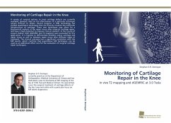
A variety of surgical options to treat cartilage defects are currently available, however, data on the effectiveness of a particular technique remain difficult to obtain. Recent progress in MRI technology has yielded quantitative techniques that can directly visualize the molecular ultrastructure of cartilage. These new techniques may allow for a biochemical analysis of the repair tissue after surgicial cartilage repair, and have a high potential to improve clinical research. In the course of several studies, both dGEMRIC and T2-mapping were evaluated for this application. It is feasible to differentiate between native cartilage and repair tissue as well as between repair tissue after different types of treatment. The clinical outcome has a significant correlation with the MR results, which confirms that quantitative MR techniques can be used as an additional effect size for the evaluation of surgical cartilage repair techniques.
Bitte wählen Sie Ihr Anliegen aus.
Rechnungen
Retourenschein anfordern
Bestellstatus
Storno

