Pellerito, John S., MD, FACR, FSRU, FAIUM (Associate Professor of Ra, Joseph F. Polak (Tufts University School of Professor of Radiology
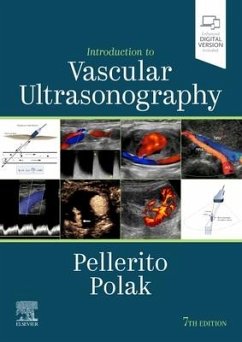
Introduction to Vascular Ultrasonography
Pellerito, John S., MD, FACR, FSRU, FAIUM (Associate Professor of Ra, Joseph F. Polak (Tufts University School of Professor of Radiology
Introduction to Vascular Ultrasonography
- Gebundenes Buch
- Merkliste
- Auf die Merkliste
- Bewerten Bewerten
- Teilen
- Produkt teilen
- Produkterinnerung
- Produkterinnerung
"Enhanced digital version included"--Cover.
Andere Kunden interessierten sich auch für
![The Vascular System The Vascular System]() Kupinski, Ann Marie, PhD, RVTThe Vascular System143,99 €
Kupinski, Ann Marie, PhD, RVTThe Vascular System143,99 €![Chapman & Nakielny's Guide to Radiological Procedures Chapman & Nakielny's Guide to Radiological Procedures]() Ravivarma Balasubramaniam (University Hospit Department of ImagingChapman & Nakielny's Guide to Radiological Procedures51,99 €
Ravivarma Balasubramaniam (University Hospit Department of ImagingChapman & Nakielny's Guide to Radiological Procedures51,99 €![Vascular Technology Examination PREP, Second Edition Vascular Technology Examination PREP, Second Edition]() Raymond GaiserVascular Technology Examination PREP, Second Edition76,99 €
Raymond GaiserVascular Technology Examination PREP, Second Edition76,99 €![Learning Radiology Learning Radiology]() William Herring (Vice Chairman and Albe Residency Program DirectorLearning Radiology60,99 €
William Herring (Vice Chairman and Albe Residency Program DirectorLearning Radiology60,99 €![Quality Management in the Imaging Sciences Quality Management in the Imaging Sciences]() Jeffrey Papp (Professor of Physics and College Diagnostic ImagingQuality Management in the Imaging Sciences90,99 €
Jeffrey Papp (Professor of Physics and College Diagnostic ImagingQuality Management in the Imaging Sciences90,99 €![MRI and Traumatic Brain Injury, An Issue of Neuroimaging Clinics of North America MRI and Traumatic Brain Injury, An Issue of Neuroimaging Clinics of North America]() MRI and Traumatic Brain Injury, An Issue of Neuroimaging Clinics of North America99,99 €
MRI and Traumatic Brain Injury, An Issue of Neuroimaging Clinics of North America99,99 €![Thoracic Imaging: Case Review Thoracic Imaging: Case Review]() Stowell, Justin T., MD (Department of Radiology, Mayo Clinic, JacksoThoracic Imaging: Case Review70,99 €
Stowell, Justin T., MD (Department of Radiology, Mayo Clinic, JacksoThoracic Imaging: Case Review70,99 €-
-
-
"Enhanced digital version included"--Cover.
Hinweis: Dieser Artikel kann nur an eine deutsche Lieferadresse ausgeliefert werden.
Hinweis: Dieser Artikel kann nur an eine deutsche Lieferadresse ausgeliefert werden.
Produktdetails
- Produktdetails
- Verlag: Elsevier - Health Sciences Division
- 7 ed
- Seitenzahl: 882
- Erscheinungstermin: 20. November 2019
- Englisch
- Abmessung: 266mm x 192mm x 32mm
- Gewicht: 1640g
- ISBN-13: 9780323428828
- ISBN-10: 0323428827
- Artikelnr.: 57909087
- Herstellerkennzeichnung
- Libri GmbH
- Europaallee 1
- 36244 Bad Hersfeld
- gpsr@libri.de
Professor, Donald and Barbara Zucker School of Medicine at Hofstra/Northwell, Department of Radiology, New York, United States.
Section 1: Basics
1. The Hemodynamics of Vascular Disease
2. The Physics and Instrumentation of Ultrasound Imaging in Vascular
Disease
3. Doppler Flow Imaging and Spectral Analysis
Section 2: Cerebral Vessels
4. Anatomy of the Cerebral Vasculature
5. Carotid Sonography: Protocols and Technical Considerations
6. Evaluating Carotid Plaque and Intima Media Thickness
7. Ultrasound Assessment of Carotid Stenosis
8. How to Assess Difficult and Uncommon Carotid Cases
9. Ultrasound Assessment of the Vertebral Arteries
10. Ultrasound Assessment of the Intracranial Arteries
Section 3: Extremity Arteries
11. Anatomy of the Upper and Lower Extremity Arteries
12. Physiologic Testing of Lower Extremity Arterial Disease
13. Assessment of Upper Extremity Arterial Disease
14. Ultrasound Evaluation Before and After Hemodialysis Access
15. Ultrasound Assessment of Lower Extremity Arteries
16. Ultrasound Assessment During and After Carotid Peripheral Intervention
17. Ultrasound in the Assessment and Management of Arterial Emergencies
Section 4: Extremity Veins
18. Extremity Venous Anatomy and Technique for US Examination
19. Ultrasound Diagnosis of Lower Extremity Venous Thrombosis
20. Risk Factors and the Role of Ultrasound in the Management of Extremity
Venous Disease
21. Diagnostic Testing for Venous Insufficiency
22. Nonvascular Findings Encountered During Venous Sonography
Section 5: Abdomen and Pelvis
23. Anatomy and Normal Doppler Signatures of Abdominal Vessels
24. Ultrasound Assessment of the Abdominal Aorta
25. Ultrasound Assessment Following Endovascular Aortic Aneurysm Repair
26. Doppler Ultrasound of the Mesenteric Vasculature
27. Ultrasound Assessment of The Hepatic Vasculature
28. Duplex Ultrasound Assessment of Native Renal Vasculature
29. Duplex Ultrasound Evaluation of the Uterus and Ovaries
30. Duplex Ultrasound Evaluation of the Male Genitalia
31. Evaluation of Organ Transplants
Section 6: Trends in Vascular Imaging
32. Credentialing, Accreditation and Quality in the Vascular Laboratory
33. Ultrasound Screening for Vascular Disease
34. Correlative Imaging in Vascular Disease
35. Ultrasound Contrast Agents in Vascular Disease

