
Broschiertes Buch
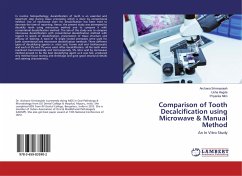
Eine In-Vitro-Studie
12. November 2024
Verlag Unser Wissen
Broschiertes Buch
An In Vitro Study
1. Februar 2016
LAP Lambert Academic Publishing
Broschiertes Buch
Uno studio in vitro
12. November 2024
Edizioni Sapienza
Broschiertes Buch
12. November 2024
Editions Notre Savoir
Broschiertes Buch
12. November 2024
Edições Nosso Conhecimento
Broschiertes Buch
12. November 2024
Ediciones Nuestro Conocimiento
Ähnliche Artikel

Broschiertes Buch
Eine CBCT-Analyse
13. September 2024
Verlag Unser Wissen
Broschiertes Buch
30. Juli 2024
Verlag Unser Wissen

Broschiertes Buch
Studie bei Kindern im Alter von 5-6, 12 und 15 Jahren aus Nord- und Süd-Kivu in der Demokratischen Republik Kongo
29. September 2024
Verlag Unser Wissen


Broschiertes Buch
Ein Einblick in die Mundhöhle
31. Januar 2024
Verlag Unser Wissen

Broschiertes Buch
Verständnis, Diagnose und Management
15. September 2024
Verlag Unser Wissen

Broschiertes Buch
Bewertung und Vergleich des Unterschieds in der Cuspaldurchbiegung zwischen verschiedenen restaurativen Kompositharzen und -techniken
20. Mai 2024
Verlag Unser Wissen

Broschiertes Buch
Ein umfassender Leitfaden zur Zahngesundheit
17. August 2023
Verlag Unser Wissen

Ähnlichkeitssuche: Fact®Finder von OMIKRON
