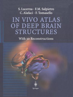
In the first half of the twentieth century, the study of neuroanatomy was essentiallybased on the observations made by scientists on brain cadavers fixed with standard techniques. These studies have produced well-known tools such as the stereotactic atlas, which have proven to be extremely useful and irreplaceable for neurosurgeons, neuroradi ologists, neurologists and neuroanatomists. In particular, the Talairach and Schaltenbrandt atlases are considered the most presti gious and up-to-date work available today. The recent introduction of neuroimaging, especially nuclear magnetic resonance, together with the exciting and tremendous progress made in computer graphics, has allowed us to approach neuroanatomy directly in living patients with more accuracy and a high degree ofdetail. This work, after a short introduction which explains the methodolo gy used, is divided into four types of sections: three types ofsections obtained from the same brain and orientated in the standard axial, sagittal, and coronal spatial planes and one type of section of three dimensional pictures obtained from the computerized processing of the previous pictures. The organization and the life-size tables obtained by magnetic reso nance make this work similar to a classic stereotactic atlas, although the authors do not claim to reach the high level of precision which such atlases usually provide. The abbreviations used are based on Latin nomenclature,in order to be understood and recognized world wide, and are supported by a system of color codes useful for the identification ofbrain structures.
Hinweis: Dieser Artikel kann nur an eine deutsche Lieferadresse ausgeliefert werden.
Hinweis: Dieser Artikel kann nur an eine deutsche Lieferadresse ausgeliefert werden.
From the reviews:
"This colour atlas of deep brain nuclei and other structures will prove to be a useful aid to both the functional neurosurgeon and the discerning neuroradiologist. ... The in vivo image capture, obtained with MRI, provides a more accurate view of these deep-seated neuroanatomical structures, making it superior to those atlases based on cadaveric imaging. Consequently, this atlas will prove useful both in the operating theatre and reporting room alike. The atlas is well organized and easy to navigate." (R.A. Trivedi, European Radiology, Vol. 14 (8), 2004)
"This is a 3D atlas of brain structures acquired from MRI of normal volunteers, produced by the Department of Neurosurgery in Messina, Italy. ... it is truly a pictorial atlas. ... The 3D images are impressive and well produced technically ... . In summary, this atlas may have most appeal to neurosurgeons but is a useful refresher for neuroradiologists ... ." (P.White, Neuroradiology, Vol. 44 (11), 2002)


