- Gebundenes Buch
- Merkliste
- Auf die Merkliste
- Bewerten Bewerten
- Teilen
- Produkt teilen
- Produkterinnerung
- Produkterinnerung
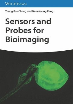
Systematically summarizes the bioimaging chemical probes from chemical points of views.
Andere Kunden interessierten sich auch für
![Intracellular Thermometry with Fluorescent Molecular Thermometers Intracellular Thermometry with Fluorescent Molecular Thermometers]() Seiichi UchiyamaIntracellular Thermometry with Fluorescent Molecular Thermometers90,99 €
Seiichi UchiyamaIntracellular Thermometry with Fluorescent Molecular Thermometers90,99 €![Point-of-Care Biosensors for Infectious Diseases Point-of-Care Biosensors for Infectious Diseases]() Point-of-Care Biosensors for Infectious Diseases105,99 €
Point-of-Care Biosensors for Infectious Diseases105,99 €![Biological Reaction Engineering Biological Reaction Engineering]() Elmar HeinzleBiological Reaction Engineering169,00 €
Elmar HeinzleBiological Reaction Engineering169,00 €![Biomedical Engineering Biomedical Engineering]() Hossein HosseinkhaniBiomedical Engineering149,00 €
Hossein HosseinkhaniBiomedical Engineering149,00 €![Mass Spectrometry for Lipidomics Mass Spectrometry for Lipidomics]() Mass Spectrometry for Lipidomics221,99 €
Mass Spectrometry for Lipidomics221,99 €![Drug Delivery Applications of Noninvasive Imaging Drug Delivery Applications of Noninvasive Imaging]() Mei TianDrug Delivery Applications of Noninvasive Imaging216,99 €
Mei TianDrug Delivery Applications of Noninvasive Imaging216,99 €![Theory and Applications of Heat Transfer in Humans, 2 Volume Set Theory and Applications of Heat Transfer in Humans, 2 Volume Set]() Theory and Applications of Heat Transfer in Humans, 2 Volume Set283,99 €
Theory and Applications of Heat Transfer in Humans, 2 Volume Set283,99 €-
-
-
Systematically summarizes the bioimaging chemical probes from chemical points of views.
Produktdetails
- Produktdetails
- Verlag: Wiley-VCH
- Artikelnr. des Verlages: 1134960 000
- 1. Auflage
- Seitenzahl: 368
- Erscheinungstermin: 2. August 2023
- Englisch
- Abmessung: 249mm x 173mm x 23mm
- Gewicht: 868g
- ISBN-13: 9783527349609
- ISBN-10: 352734960X
- Artikelnr.: 67395202
- Herstellerkennzeichnung
- Wiley-VCH GmbH
- Boschstraße 12
- 69469 Weinheim
- wiley.buha@zeitfracht.de
- 06201 6060
Young-Tae Chang studied chemistry in Pohang University of Science and Technology (POSTECH, Korea) and received his B.S. in 1991. After one and half years of army service in Korea, he started his graduate study at POSTECH and received a Ph.D. in 1997 under the supervision of Prof. Sung-Kee Chung, working on the divergent synthesis of all possible regioisomers of myo-inositol phosphates. He did his postdoctoral work with Prof. Peter Schultz at UC Berkeley and The Scripps Research Institute. In 2000, he was appointed assistant professor at New York University and promoted to associated professor in 2005. In September, 2007, he moved to National University of Singapore and Singapore Bioimaging Consortium. He was a full professor of Chemistry and leader of Medicinal Chemistry Program of NUS, and Lab Head of Bioimaging Probe Development at SBIC, Biopolis. In 2017, he came back to POSTECH, as a professor in the Department of Chemistry, and joined Center for Self-assembly and Complexity (CSC), Institute for Basic Science (IBS) as an Associate Director. He published more than 300 scientific papers / 2 books and filed more than 50 patents so far. Nam-Young KANG is a Research Professor at the Department of Convergence IT Engineering, Pohang University of Science and Technology (POSTECH). Her research of interest is focused on Cell image-based High contents/through-put screening (HCS/HTS) to develop HCS based new drug using fluorescent imaging probes.
1 INTRODUCTION OF BIOIMAGING
1.1. Color
1.2 Colorful material
1.3 Light source of bioimaging
1.4 Subcellular imaging
1.5 Cell selective imaging
1.6 Tissue and organ imaging
1.7 Whole body imaging
1.8 Probes in bioimaging
2. CHEMICAL SENSORS AND PROBES FOR BIOIMAGING
2.1 History of dyes in biological stains
2.2 Blood cell staining
2.3 Bacteria staining using Gram method
2.4 Fluorescent sensors and probes
2.5 Representative fluorescent compounds for bioimaging
3. ORGANELLE SELECTIVE PROBES
3.1 Cell plasma membrane
3.2 Endosome and lysosome
3.3 Nucleus and DNA
3.4 Nucleolus and RNA
3.5 ER and Golgi body
3.6 Mitochondria
3.7 Lipid droplet
3.8 Peroxisome
3.9 Cytosol
3.10 Extracellular vesicle
3.11 Nonmembrane-bound condensate
3.12 Organelle probes in live cells and fixed cells
3.13 Modeling for organelle selective probes
4. LIVE CELL SELECTIVE PROBES
4.1. Protein Oriented Live Cell Distinction (POLD)
4.2. Carbohydrate Oriented Live Cell Distinction (COLD)
4.3. Lipid Oriented Live Cell Distinction (LOLD)
4.4. Gating Oriented Live Cell Distinction (GOLD)
4.5. Metabolism Oriented Live Cell Distinction (MOLD)
5. EX-VIVO TISSUE SELECTIVE PROBES
5.1 Immunohistochemistry
5.2 Tissue imaging with nucleic acid probes
5.3 Tissue imaging with small molecule probes
5.3.1 Pancreatic islet imaging
5.3.2 Neuronal tissue imaging
5.4 Organoid as model of tissue and organ
6. IN-VIVO WHOLE-BODY IMAGING PROBES
6.1 ElaNIR for elastin imaging in mouse
6.2 Probes for exposed neuron in zebrafish embryo
6.3 NeuO for whole-body neuron imaging in zebrafish
6.4 LipidGreen for fatty tissue imaging in zebrafish
6.5 Blood vessel imaging in zebrafish
6.6 Probes for bone imaging
6.7 Probes for pancreatic islet imaging
6.8 Probes for eye imaging
6.9 Macrophage imaging in ischemia and inflammation
7. IMAGING FOR BIOLOGICAL ENVIRONMENT AND FUNCTION
7.1 pH
7.2 Metal ions
7.3 Metabolites
7.4 Viscosity
7.5 Temperature
7.6 ROS and RNS
8. IMAGING FOR DISEASE
8.1 Cancer imaging
8.2 Neurodegenerative disease imaging
8.3 Inflammation imaging
8.4 Diabetes imaging
8.5 Liver disease imaging
8.6 Aging
8.7 Theranostics
9. NON-OPTICAL IMAGING PROBES
9.1 Ultrasound imaging probes
9.2 X-ray contrast agents
9.3 MRI contrast agents
9.4 SPECT probes
9.5 PET probes
9.6 Multimodality
10. FLUORESCENCE IMAGING TECHNIQUES AND ANALYSIS METHODS
10.1 Multi-color imaging
10.2 Ratiometric measurement
10.3 Fluorescence lifetime imaging microscopy
10.4 Confocal fluorescence microscopy
10.5 Two photon excitation fluorescence imaging and harmonic generation
10.6 Selective plane illumination microscopy
10.7 Total internal reflection fluorescence microscopy
10.8 Super resolution imaging
10.9 Single molecule imaging
10.10 Photoactivation of caged molecule
10.11 Fluorescence recovery after photobleaching
10.12 Flow cytometry technique
11. PERSPECTIVES FOR FUTURE PROBE DEVELOPMENT
11.1 Design of selective probes
11.2 Discovery of selective probes by screening
11.3 Future probe development

